Growth and Cyclical Changes in the Reproductive Organs
At midgestation the fetal ovaries are much larger than those of the dam but they later regress by birth to one-tenth of their greatest fetal size. They then grow slowly until puberty when a sudden spurt occurs.
The first estrus is generally at the beginning of a breeding season, and the age of first estrus varies with the date of the individual's birth as well as with breed and nutrition. It usually occurs sometime between the 18th and 27th months. The neonatal ovary is ellipsoidal but develops into the peculiar indented adult form during the first 2 or 3 years (Fig. 22.15). In the mature ovary the larger follicles are concentrated near the ovulation fossa to which they migrate as they enlarge (Fig. 22.10/2). Two or three (perhaps spread between the two ovaries) reach full size of about 5 cm in each cycle, but usually only one ruptures. After rupture, the cavity contains some blood, and for a time the soft clot may be appreciated on rectal examination. It then gradually fills with luteal cells, but even when mature the corpus luteum hardly projects above the surrounding surface. The corpus luteum is initially brick red but becomes ocherous as it matures. Its regression begins about the 10th day and is more or less complete when its successor forms. The cycle averages 22 days. The left ovary is generally the more active; despite this the right uterine horn is slightly more favored by conceptuses. Transuterine migration by a conceptus must be common.Ultrasonography may be used to follow follicular development, to detect the occurrence of ovulation, and to trace the fate of the resulting follicular cavity. It successfully determines the course of events a little before this is possible by palpation per rectum. It may allow the prediction of ovulation by about a day because it can reveal the change in form, from spherical to pyriform, of the ripening follicle.
A further advantage lies in its success in recognizing the parallel maturation of multiple follicles that may result in twin pregnancy.The juvenile reproductive tract is small, symmetrical, and thin walled. The endometrium is pale, and the layers of the uterine wall are difficult to differentiate with the naked eye. The broad ligaments are thin and transparent, and the blood vessels are narrow and relatively inconspicuous. Growth is initially isometric—it keeps pace with growth of the body as a whole—until a prepubertal acceleration occurs. Cyclical changes in the uterus, including increased retention of water, a greater blood flow, and activation of the glands thickening the wall in preparation for the reception of the blastocyst, broadly resemble those in other species. If pregnancy does not result, these changes recede with the regression of the corpus luteum. Cyclical changes in muscular tonus are the subject of some controversy, but most authorities hold that tonus is greatest about a week after ovulation.
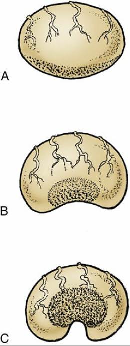
FIG. 22.15 The postnatal development of the ovary. The more rapid growth at the poles confines the germinal epithelium (stippled) to a small central area. (A) At birth; the germinal epithelium is widespread over the surface. (B) At 6 months of age. (C) Adult; the germinal epithelium surrounds an indentation known as the ovulation fossa.
The cervix softens during estrus when the intravaginal part droops so that its orifice is lost to view on vaginoscopic examination (Fig. 22.12B). When stimulated by handling, it becomes firmer, returns to the horizontal, and may exhibit rhythmic contractions. It is also moist, swollen, and pink at this time. It is paler in appearance and firmer during metestrus and diestrus when the lumen is closed by a plug of thick mucus (Fig. 22.12). Although the vaginal wall is pink and moist during estrus, its liability to change color on prolonged exposure to air denies diagnostic significance to its appearance. Cytologic changes in the vaginal epithelium are slight and also of little diagnostic value.