Hearing and Balance
The ear can be divided into three main parts: the external, middle, and inner ears. The external ear extends from the exterior as far as the tympanic membrane (eardrum). The middle ear begins at the tympanic membrane; it is an air-filled space within the temporal bone.
The inner ear is housed entirely in the temporal bone, forming an elaborate fluid-filled system of chambers and canals (Fig. 11-6).External Ear
The part of the ear visible on the outside of the head, the auricle, or pinna, varies considerably in shape and size between and within species. its appearance is dictated primarily by the shape and rigidity of the auricular cartilage, an elastic cartilage that is somewhat funnel shaped. The pinna acts as a capturing device for air pressure waves, and its shape and mobility are structurally important for sound localization. The many skeletal muscles that move the pinna are loosely categorized as rostral and caudal auricular muscles.
A tubular extension of the pinna, the cartilaginous external acoustic canal, extends from the pinna to the tympanic membrane. The external acoustic canal is lined with a modified skin possessing few hairs and abundant sebaceous and ceruminous glands. The combined secretion of these glands is called cerumen, a brown waxy substance that protects the canal. A separate annular cartilage adjacent to the funnel-shaped base of the auricular cartilage forms most of the external acoustic canal. A third cartilage, the scutiform cartilage, lies medial to the auricular cartilage, embedded in the rostral auricular muscles. it functions as a fulcrum for the attached auricular muscles.
Middle Ear
The middle ear is an air-filled space, the tympanic cavity, lined by mucous membrane and contained within the temporal bone. in most domestic animals, the middle ear features a ventrally expanded cavity, the tympanic bulla, visible on the ventral surface of the skull.
The middle ear is closed to the external acoustic canal by the intact tympanic membrane and communicates with the nasopharynx via the auditory tube (formerly eustachian tube). in the horse, the auditory tube is expanded to form the large air-filled guttural pouch dorso- caudal to the nasopharynx. The connection between the middle ear and the pharynx is normally closed except briefly during swallowing. The opening of the auditory tube brought about by swallowing or yawning permits equalization of air pressures between middle and external ears.Three auditory ossicles span the middle ear from tympanic membrane to the vestibular (oval) window. From superficial to deep, they are the malleus (hammer), incus (anvil), and stapes (stirrup) (Fig. 11-6). These tiny bones provide a mechanical linkage from the tympanic membrane to the vestibular window, and through them vibrations are transmitted from the former to the latter. The size and leverage of the auditory ossicles provide mechanical advantage; the energy of pressure waves striking the tympanic membrane is concentrated on the smaller vestibular window, increasing the system’s sensitivity to faint stimuli.
There are also two striated muscles within the middle ear, the m. tensor tympani and the
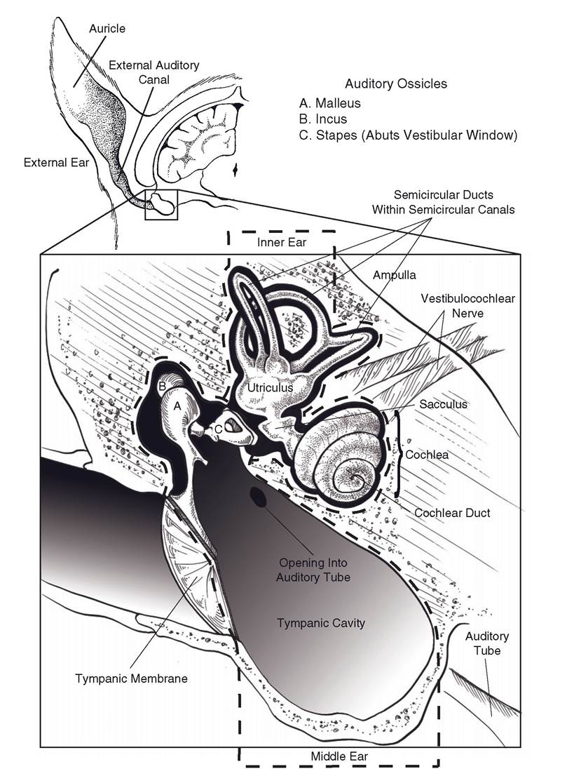
Figure 11-6. Anatomy of the hearing and vestibular apparatus. External, middle, and internal ears with surrounding temporal bone.
m. stapedius. These two small muscles damp vibrations of the auditory ossicles in the presence of excessively loud noises. It is persistent contraction of these muscles that contributes to the temporarily reduced hearing acuity notable after, for example, attending a very loud music concert.
The chorda tympani, a branch of the facial nerve, passes through the tympanic cavity. The chorda tympani carries fibers for taste from the rostral two-thirds of the tongue and also parasympathetic fibers destined for the mandibular and sublingual salivary glands.
infections of the middle ear (otitis media) can be associated with dysfunction of this nerve. Likewise, sympathetic fibers that innervate the eye pass through the tympanic cavity; diseases affecting the middle ear can produce signs of lost sympathetic innervation to head structures (Horner’s syndrome), including a constricted pupil, sunken globe, and drooping of the upper eyelid.In horses, cranial nerves VII and IX through XII and the continuation of the sympathetic trunk pass adjacent to the guttural pouch, separated from its interior only by mucous membrane. Diseases of the guttural pouch can therefore produce distinctive neurologic dysfunctions when these nerves are affected.
Internal Ear
The internal ear is housed entirely within the petrous temporal bone. it is a multichambered membranous sac (the membranous labyrinth) closely surrounded by a sculpted cavity of bone (the osseous labyrinth). The inner ear detects both sound and acceleration of the head (Fig. 11-6).
The membranous labyrinth in the internal ear is a system of fluid-filled sacs and ducts. The primary anatomic features of the membranous labyrinth are (1) two enlargements, the utriculus (utricle) and sacculus (saccule); (2) three loops attached to the utriculus, the semicircular ducts; and (3) the spiraled cochlear duct. These are filled with a fluid, the endolymph. The membranous labyrinth is housed in the osseous labyrinth, a similarly shaped, slightly larger excavation in the petrous temporal bone. The osseous labyrinth is filled with perilymph.
The inner ear may be divided into two functional parts. in one, the osseous labyrinth forms a coiled, snail shell-shaped cochlea, inside of which is the cochlear duct, an extension of the membranous labyrinth. The cochlear duct houses the receptors for audition. These receptors are innervated by the cochlear division of the vestibulocochlear nerve.
The second part constitutes the vestibular apparatus. Within the utriculus, sacculus, and semicircular ducts that constitute the vestibular apparatus are the receptors that detect accelerations of the head, including acceleration due to gravity.
Accelerations of the head contribute to the sense of equilibrium or balance. The vestibular apparatus is innervated by the vestibular division of the vestibulocochlear nerve.Physiology of Hearing
The environmental energy detected in audition is air pressure waves produced by vibration. These pressure waves can be described in terms of their frequency, the time between peaks of pressure waves, measured in hertz (Hz, cycles per second). Frequency determines the perceived pitch of sounds, with higher frequencies producing sounds of higher pitches. Air pressure waves are also described in terms of their amplitude, a property that reflects the energy and consequently the loudness of these waves. Amplitude is expressed in decibels (dB), the units by which loudness is measured. The decibel scale is logarithmic, so that the loudest sounds that can be heard without discomfort (around 100 dB) are a million times as energetic as the faintest audible sounds.
The cochlear portion of the osseous labyrinth resembles a snail shell (cochlea is Latin for snail). The space on the inside of the cochlea is full of perilymph, and it spirals around a central bony core, the modiolus. The corresponding part of the membranous labyrinth is the cochlear duct, which extends throughout the coiled length of the cochlea. The duct is stretched transversely from the modiolus to the outer wall of the bony cochlea, effectively dividing this perilymph-filled space into two: the scala vestibuli above and the scala tympani below the duct.
The scala vestibuli originates in the region of the vestibular window, and by this association, the perilymph within it receives pressure waves from the vibration of the auditory ossicles. At the apex of the cochlea the scala vestibuli is continuous with the scala tympani at a connection called the helicotrema. The scala tympani receives pressure waves from the fluid in the scala vestibuli (but more importantly, through vibrations transmitted through the intervening cochlear duct); these waves are dissipated at the termination of the scala tympani, the cochlear (round) window, which abuts the air-filled space of the middle ear.
The receptor cells of the auditory system are within the cochlear duct as components of the spiral organ (organ of Corti) (Fig. 11-7). The spiral organ contains the receptor cells of the internal ear, mechanoreceptors called hair cells for the bundle of cilia on their apex. The cilia of the hair cells in the spiral organ are embedded in a relatively stiff overlying membrane, the tectorial membrane. The walls between the cochlear duct and the scalae vestibuli and tympani are called the vestibular and basilar membranes, respectively.
Cross-sectional views of the cochlea give the impression that the components within are arrayed as repeating separate units, but they are longitudinally continuous throughout the extent of the spiraled cochlea.
The hair cells synapse with peripheral processes of primary afferent neurons whose cell bodies lie within the spiral ganglion. The spiral ganglion is housed in the modiolus, and the axons of the afferent neurons within it gather to form the cochlear nerve. Transduction of the mechanical energy of air pressure waves into the electrical impulses of neurons in the auditory system takes place in the following manner.
Air pressure waves captured by the pinna are funneled down to the tympanic membrane,
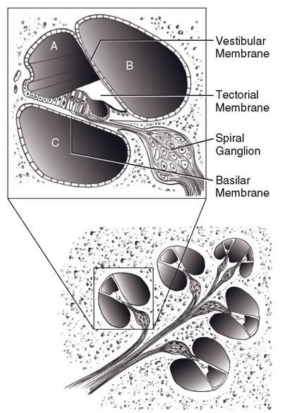
Figure 11-7. Median section of the cochlea. Expanded view shows spiral organ within: A, cochlear duct; B, between the scala vestibuli; and C, scala tympani.
and they put that membrane into vibratory motion. This motion is carried across the airfilled cavity of the middle ear by movements of the auditory ossicles. These ossicles transfer the vibrations to the vestibular window via the foot of the stapes. The perilymph in the osseous labyrinth receives the pressure waves transmitted to it by the vibrations of the vestibular window. These are carried into the cochlea by the scala vestibuli. The vestibular membrane between scala vestibuli and cochlear duct vibrates in response to the pressure waves and transfers those vibrations across the cochlear duct to the basilar membrane and underlying perilymph of the scala tympani. Movement of the basilar membrane results in movement of the hair cells in the spiral organ that rests upon it, and this produces bending of the hair cells’ cilia against the more rigid tectorial membrane.
It is the bending of the cilia that results in a depolarization within the hair cells. When this depolarization reaches sufficient intensity, it initiates an action potential in the primary affer- ents of the spiral ganglion.The ability to discriminate one pitch from another has its basis in a number of anatomic and electrical features of the spiral organ. Most simply, the basilar membrane varies in width from the base of the cochlear duct, where it is most narrow, to the apex, where it is widest. This difference in width means that each portion of the membrane has a different frequency at which it preferentially vibrates, with the widest part of the membrane vibrating at low frequency and the narrow part at high.
The axons of the cochlear nerve enter the brainstem with the vestibular nerve at the junction of the medulla and pons and terminate in cochlear nuclei on the lateral side of the medulla. From this first synapse, multiple pathways produce auditory-mediated reflexes and conscious perception of sound. Auditory information destined for the cortex ascends bilaterally in the medulla, pons, and midbrain. The pathways for conscious perception of sound continue on to the thalamic relay nucleus for audition, and from there fibers project to the primary auditory cortex on the lateral aspect of the cerebrum. The result of bilaterality of the pathways is that auditory information from both cochleae reaches both left and right auditory cortices.
The bilaterality of auditory representations in the brain means that for a brain lesion to produce complete deafness, it must affect both sides of the pathway. Such brain injuries are often incompatible with life; therefore, most deafness is peripheral (associated with the cochlear nerve or internal ear rather than the brain itself). In animals, incomplete loss of hearing is difficult to detect.
Disease processes that affect the ability of the tympanic membrane or auditory ossicles to transmit vibrations to the vestibular window produce conduction deafness. Those that affect the spiral organ or more proximal components of the auditory system (including the cochlear nerves, brainstem, and auditory cortices) produce sensorineural deafness. Most inherited deafness is sensorineural, brought about by degeneration of cochlear hair cells. Congenital deafness has been associated with white hair coats or the merle or piebald color genes in a variety of species, including dogs (most notably the Dalmatian), cats, and horses. In these individuals, lack of pigment-containing cells inside the cochleae appears to be linked to degeneration of cochlear hair cells within a few weeks of birth.
Mechanisms of Balance
The vestibular system is a complex neurologic system that is concerned with maintaining a stable orientation in relation to gravity and while in motion. its influence is widely distributed throughout the nervous system; vestibular input is responsible for the reflex position of eyes, neck, trunk, and limbs in reference to movement or position of the head.
The receptor organs of the vestibular system are housed in the part of the membranous labyrinth known as the vestibular apparatus (Fig. 11-6). These receptor organs are the maculae of the utriculus and sacculus and the cristae ampullares of the semicircular ducts. Afferent information from these structures gives rise to motor reflexes that maintain stabile visual images on the retinae during movement of the head, to keep the head level with respect to gravity through neck movements, and to produce trunk and limb movements to counteract displacements of the head.
The utriculus and the smaller sacculus each posses a macula, a thickened oval plaque of neuroepithelium (Fig. 11-8A). The maculae consist of a population of hair cells very like those of the spiral organ. These are covered by a gelatinous sheet, the otolithic membrane, into which project the cilia of the hair cells. The surface of the otolithic membrane is studded with crystals of calcium carbonate, the otoliths, or statoconia, which increase the inertial mass of the otolithic membrane. When the head accelerates in a straight line, the individual
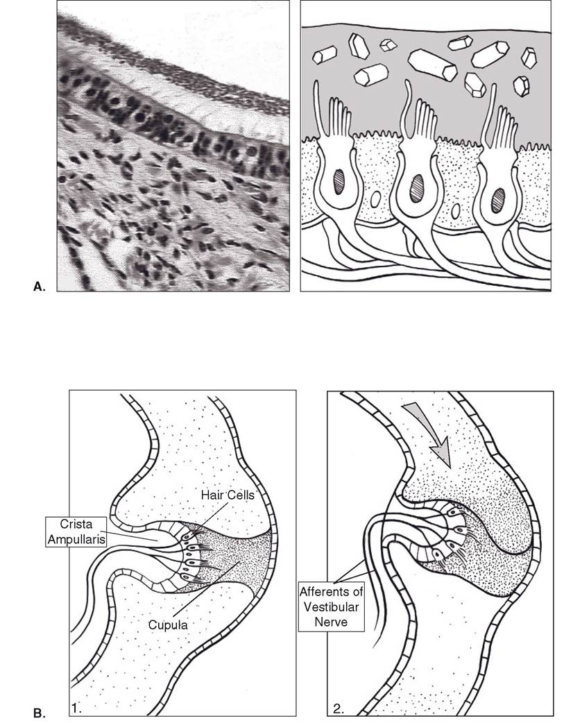
Figure 11-8. Receptor organs of the vestibular apparatus. A) Anatomy of the macula. Hair cells, surrounded by nonneural supportive cells, are surmounted by a gelatinous otolithic membrane in which are embedded calcium carbonate crystals called otoliths. Inertial movements of the otolithic membrane bend the cilia of the hair cells, changing their membrane potential. Inset shows photomicrograph of a macula. B) 1. Anatomy of the crista ampullaris. Hair cells and supportive cells are found in a crest of tissue on one side of the ampulla. 2. A gelatinous cupula, in which the hair cells are embedded, forms a flexible barrier across the ampulla. Inertial movements of endolymph bend the cupula, bending the cilia of the hair cells.
senses linear acceleration. The inertia of the otolithic membrane causes it to lag behind the head under conditions of linear acceleration (including the always present acceleration due to gravity); this dragging of the otolithic membrane bends the cilia of the underlying hair cells with shearing vectors dependent on the direction of acceleration.
Attached to the utriculus are three halfcircular extensions of the membranous labyrinth, the semicircular ducts. The ducts lie in three planes at approximately right angles to one another and are designated anterior, posterior, and lateral to describe their orientation. As an extension of the membranous labyrinth, each duct is filled with endolymph and surrounded by perilymph.
one end of each duct is dilated to form an ampulla, within which are housed the receptor organs of the semicircular ducts (Fig. 11-8B). one wall of the ampulla features a transverse ridge of connective tissue, the crista ampullaris, which supports a neuroepithelium of hair cells. Attached to the crista is a gelatinous cupula; this extends across the ampulla, forming a flexible barrier to the flow of endolymph. The cilia of the hair cells are embedded in the cupula and are therefore bent by movements of it.
The semicircular ducts detect angular acceleration (rotation), and their planes of orientation roughly correspond to the X-, Y-, and Z-axes of three-dimensional space. When the head rotates, the semicircular duct lying in that rotational plane moves with it. The endolymph inside the duct, however, must overcome its inertia at the start of rotation and, as a consequence, briefly lags behind the movement of the head. The flexible cupula, acting as a dam across the ampulla, bulges in response to the endolymph’s push, and in doing so, bends the cilia of the embedded hair cells. With three pairs (right and left) of semicircular ducts detecting movement in the three planes of space, complex rotational movements of the head are encoded in the firing patterns of the six cristae ampullares.
Primary afferent neurons synapse with the hair cells of the vestibular apparatus. Their cell bodies are in the vestibular ganglion, and their axons constitute the vestibular nerve, which joins the cochlear nerve to become the eighth cranial nerve. Most axons in the vestibular nerve synapse in the large vestibular nuclei of the pons and rostral medulla.
Vestibular Reflexes. If there were no mechanism to keep the eyes fixed on a target when the head moved, visual images would continually slip across the retina during head movement, and focusing on the visual field would be difficult or impossible while the head was moving. The vestibular nuclei use information about acceleration to coordinate extraocular muscle movements with movements of the head and thereby fix the visual image in one place on the retina as long as possible. When the excursion of the moving head carries the fixed image out of visual range, the eyes dart ahead in the direction of movement to fix upon a new image. This new image is held on the retina while the head continues turning until a compensatory jump ahead is again needed. This mechanism results in a cycle of slow movement opposite the direction of turn (eyes fixed on target) followed by a rapid readjustment in the same direction as the turn. This oscillatory reflex eye movement is called nystagmus.
Nystagmus is a normal reflex, generated in response to movement of the head. Nystagmus is considered abnormal when it occurs in absence of head movement; this resting (spontaneous) nystagmus is a sign of vestibular disease.
Axons of some neurons in the vestibular nuclei project caudad in a motor tract that influences activity in cervical and upper thoracic spinal cord segments. These motor connections activate neck musculature and forelimb extensors, producing the vestibulocollic reflex, which generates neck movements and forelimb extension to help keep the head level with respect to gravity and movement.
some fibers from the vestibular nuclei form an ipsilateral descending motor tract that extends the length of the spinal cord. This lateral vestibulospinal tract is part of the ventromedial motor system (see Chapter 9). Its activity has the primary effect of increasing tone in antigravity muscles (proximal limb extensors and axial muscles). This vestibulospinal reflex uses vestibular information to produce limb and trunk movements that counteract the displacement of the head that elicits it. This mechanism is designed to prevent tilting or falling with shifts in head position.
Animals and people with injury to one side of their vestibular system express the vestibular reflexes in the absence of appropriate stimuli. These individuals commonly have resting nystagmus, a head tilt, and a tendency to lean, circle, or fall to one side.