Hydrocephalus Is an Increased Volume of Cerebrospinal Fluid in the Skull
Hydrocephalus is defined as an increased CSF volume in the skull, often associated with an increased ventricular volume and increased intracranial pressure. In theory, hydrocephalus could be caused by too much fluid production at the choroid plexuses, obstruction to its flow through the ventricular system or subarachnoid space, or impaired absorption at the arachnoid villi.
In practice, overproduction seems rare, whereas obstruction to flow seems more common, particularly at such vulnerable sites as the narrow cerebral aqueduct (connecting the third and fourth ventricles) and the exits from the fourth ventricle. Such blockages in the ventricular system produce a Honconinumtcathig Iiydroeephalus that results in a buildup of freshly produced CSF in portions of the ventricular system behind the blockage. This causes the ventricular regions inside the brain to expand at the expense of the surrounding brain tissue, and intracranial pressure rises.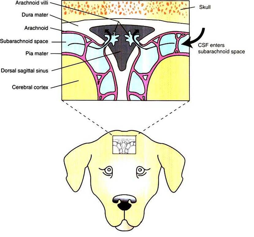
FIGURE 15-4 Transverse (coronal) section through the dorsal midline of the brain showing the absorption of CSF into the dorsal sagittal sinus, through arachnoid villi. The CSF is colored light blue. The small window on the dog's head shows the approximate dorsoventral position of the sinus. (Modified from Oliver JE, Lorenz MD: Handbook of veterinary neurology, ed 2, Philadelphia, 1993, Elsevier- Saunders,)
Impairment of absorption (a type of Connnunieating hydrocephalus) can be secondary to meningitis or hemorrhage, presumably as a result of cellular debris that obstructs the transfer of CSF from subarachnoid space to venous sinus at the arachnoid villi. This can increase CSF volume in the subarachnoid space, which increases pressure on the outside surface of the brain and increases intracranial pressure.
The pathogenesis of many cases of hydrocephalus is not known. A common form of treatment in humans is surgical implantation of a tube that shunts CSF into the atria of the heart or into the peritoneal cavity, thus relieving episodes of increased CSF pressure and preventing further brain damage.