Permeability Barriers Exist Between Blood and Brain
Many dyes, once injected into the blood, stain other tissues of the body but not the brain. This suggests that the brain’s blood vessels have the ability to restrict certain substances from accessing brain tissue.
This physiological property of CNS blood vessels is referred to as the blood-brain barrier (BBB). The BBB contributes to a stable environment for the neurons and glial cells of the CNS. As Claude Bernard noted more than a century ago, homeostasis, or constancy of the internal environment, is a precondition for autonomous life. It is not surprising that the brain, the “master choreographer of autonomous life," should have special mechanisms for protecting its internal environment. Such protection from direct exposure to the blood supply is necessary because the composition of blood can significantly vary with factors such as diet, exercise, metabolic activity, illness, age, and exposure to environmental toxins. Many of the varying blood-borne nutrients, metabolites, and toxins are neuroactive and capable of affecting membrane receptors, transporters, or ion channels. In the absence of a BBB, these substances could result in unregulated and undesirable changes in neural activity and behavior.In most capillaries, water-soluble compounds leave through open clefts between capillary endothelial cells, and exchange is relatively unrestricted. In brain capillaries, however, passage through intercellular clefts is blocked by tight junctions, and exchange of blood solutes is highly selective (Figure 15-5). As a general rule, molecules that are small, uncharged, lipid soluble, and unbound to plasma proteins (e.g., O2, CO2, ethanol, nicotine) can easily pass across the capillary endothelium of the BBB. Some molecules that do not fit this profile (e.g., glucose, some amino acids) are able to pass through the BBB by specific, carrier-mediated transport mechanisms.
Brain capillaries have a greater number of mitochondria, which reflects the operation of such transporters. Certain degrada- tive enzymes expressed by brain capillary endothelium (e.g., monoamine oxidase) provide a further restriction on substances that can pass the BBB. A notable feature of brain capillaries is that they arc surrounded by a layer of glial astrocytic “end-feet.” These end-feet arc thought to play a role in formation of the capillary endothelial tight junctions during development. Evidence also indicates that the end-feet interact with the BBB-forming capillary endothelial cells and neurons to facilitate molecular homeostasis of the brain.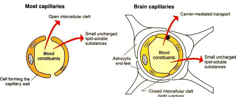
FIGURE 15-5 Blood-brain barrier (BBB). Unlike most capillaries of the body, cells of brain capillary walls are joined by tight junctions that restrict the passage of material between the cells. Materials leaving brain capillaries must pass through the cells forming the capillary wall. Substances that are not small, uncharged, and lipophilic must be carried across the cells by selective transport mechanisms. Astrocytic end-feet are thought to contribute to the tight-junction organization during development.
Unfortunately, for many patients, the protection afforded to the CNS by the BBB often prevents many antibiotics and other drugs from reaching the brain, particularly drugs with low lipid solubility or drugs bound to plasma proteins. Attempts to circumvent this problem have focused on temporary disruption of the BBB, direct delivery to the brain, “hitching a ride” on BBB membrane transporters, and increasing the lipid solubility of drugs.
In some parts of the brain known as the Circimivenfricidar organs, which include the hypothalamus, the brain capillaries do not form tight junctions, and the BBB is apparently not effective. This is significant because these brain regions are involved in functions such as the control of scrum osmolality and glucose levels, hormonal communication, feeding, drinking, and vomiting, and therefore they need to detect the levels of many serum solutes.
cranial pressure, which is reflected in an increased CSF pressure in the cisterna magna. In measuring this pressure, the dog is anesthetized, and a Styletted spinal needle is placed in the cisterna magna. The stylet is removed, and a rigid glass or plastic tube (manometer) is attached using a right-angle, three-way valve. CSF rises up the manometer to a height proportional to intracranial pressure. Its height is measured off the millimeter graduations marked on the tube.
The proprioceptive deficits of the right front and right rear legs result from a focal, asymmetric lesion of the left cerebral cortex. The seizure also resulted from this mass. With the mammary mass, metastatic lesions in the lungs, asymmetric neurological signs, seizures, and elevated CSF pressure, it is reasonable to conclude that this dog has an intracranial neoplasm that probably spread from the mammary gland to the lungs and the brain. Computed tomography (CT) or magnetic resonance imaging (MRI) is warranted to further define the tumor in the brain.
Treatment. If this dog had a focal tumor within the CNS, other treatments would be possible. However, extensive treatment in this case would be futile because of the metastatic lesions. Palliative care, steroids, and analgesics will make the animal more comfortable.