An Independent, Intrinsic Enteric Nervous System LiesWithin the Wall of the Gut
The enteric nervous system (ENS) is extensive and highly sophisticated, containing about as many neurons as the spinal cord. The ENS consists of cell bodies and their associated neurons, all of which lie within the gut wall.
Anatomical characteristics of the GI wall are illustrated in Figure 27-2, which depicts the small intestine as an example. Within the gut wall, cell bodies of the ENS are arranged into two systems of ganglia: the myenteric (Auerbach) plexus and the submucosal (Meissner) plexus. (See Chapter 13 for a more general discussion of ganglia.) The myenteric plexus consists of ganglia located between the circular and longitudinal muscle layers. The submucosal plexus has its ganglia in the submucosal layer. Axons from the cell bodies project in rich networks near the ganglia. A thick net of neurons runs in the plane between the circular and longitudinal muscle layers, connecting the ganglia of the myenteric plexus. Individual neurons leave the neuronal network to innervate structures within the gut wall and to intercommunicate between the myenteric and submucosal plexuses (Figure 27-3). Interneuronal connections within the myenteric plexus are extensive and traverse long segments of gut, whereas interneuronal connections are limited within the submucosal plexus. The intricacy of the ENS has earned it the name aIittIe brain” within the gut.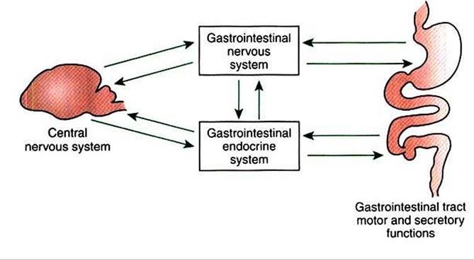
FIGURE 27-1 Gut function is under direct regulation of the enteric nervous system (ENS) and the gastrointestinal (GD endocrine system. Most central nervous system influence on the gut is mediated through indirect effects on the ENS and Gl endocrine systems.
More on the topic An Independent, Intrinsic Enteric Nervous System LiesWithin the Wall of the Gut:
-
Veterinarian -