INTEGUMENT
Avian skin is very thin as it is protected by the plumage and helps to reduce weight (Spearman 1971) (Fig. 6.71). It is lightly attached to underlying muscle but firmly attached to bone.
There are only three glands: the uropygial or preen gland, the aural gland and the vent gland. The absence of sweat glands means birds have to lose heat through their skin and by evaporation from the respiratory system.The epidermis consists of the superficial stratum corneum, which contains keratinized dead cells and the deeper living stratum germinativum. This layer is thin and fragile under the feathers but thicker on the feet and around the beak in order to resist mechanical stress. A unique feature of the avian epidermis is that it acts like a holocrine sebaceous gland, secreting a thin lipid film that helps in the maintenance of the plumage (Spearman 1983; Spearman & Hardy 1985).
The dermis is composed of connective tissue and contains the feather follicles, nerves and blood vessels. In some species, like the domestic fowl, the dermis becomes thickened and highly vascularized to form combs and wattles, but the epidermis remains thin, making them prone to injury (Dyce et al. 2002). The subcutaneous layer is mainly composed of loose connective tissue and some adipose tissue. It is here that fat is laid down in aquatic species like ducks, geese, and swans, and prior to migration in migratory species (King & McLelland 1984).
CLINICAL NOTE
Psittacines with beak malocclusion may need the lower beak trimmed, along with the upper, in order to maximize function of the beak tip organ to aid in food prehension.
Avian skin is very thin and, owing to the scant subcutaneous tissue, very inelastic. Handle tissues with care when suturing and avoid excess skin tension as it tears easily. Owing to the fact that it has less blood and a smaller nerve supply than mammals, skin wounds bleed less and are less sensitive.
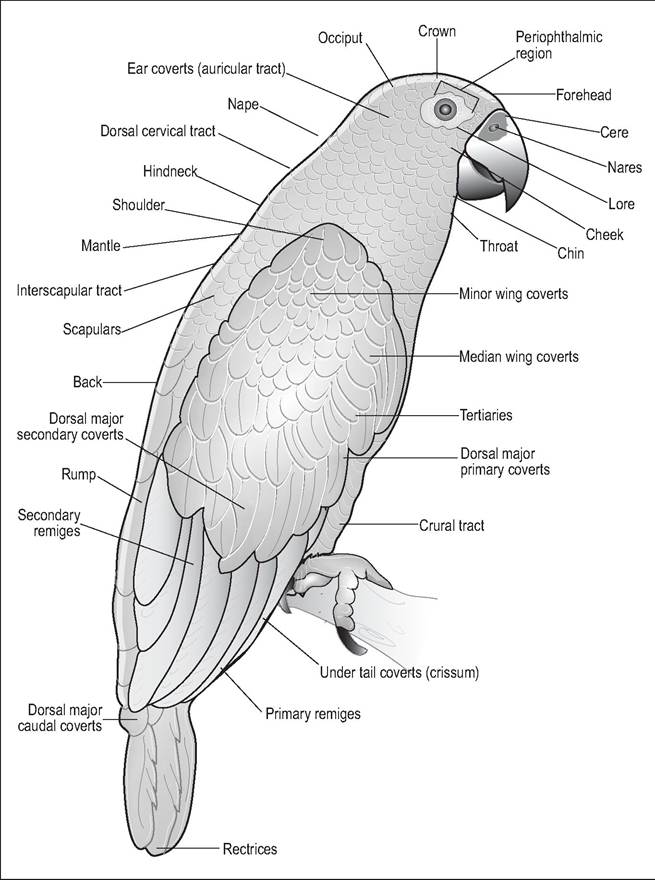
Figure 6.71 • Avian plumage.
Uropygial gland
The uropygial gland is the preen or oil gland and is found at the dorsal base of the tail (Evans 1996) (Figs. 6.72 and 6.73). It is most developed in waterfowl and maintains feather condition and waterproofing. It also acts as a bacteriostat (Spearman 1983). It is a bilobed holocrine gland drained by a papilla dorsocaudally and is covered by a tuft of down feathers called the uropygial wick. This feather tuft may aid in transmitting oil from the gland to the beak while preening (Dyce et al. 2002; Spearman & Hardy 1985). This gland is prominent in African Grays and budgies but absent in many parrots (e.g., Amazon parrots), ostriches and many pigeons (Bauck, Orosz & Dorrestein 1997; Bezuidenhout 1999; Evans 1996; Spearman 1971).
Lipid secreted by this gland and the epidermal cells are spread over the feathers by the bill during preening (Spearman 1983). This lipid layer forms a protective bacteriostatic layer over the skin and may explain why birds are less prone to skin infections. Birds can preen as often as once an hour at rest (Bauck, Orosz & Dorrestein 1997).
Aural sebaceous glands around the external ear secrete a waxy substance. Vent glands secrete mucus but their function is unknown, although it may be linked to internal fertilization.
Podotheca
The non-feathered area of the legs is called the podotheca and is composed of keratinized epidermal plates called scales. The skin is thickened in the ventral metatarsophalangeal region and is designed to withstand impact on landing. In aquatic species, the skin is softer and more flexible and modified between the toes into webs. The distal phalanx is keratinized into the nail or claw. The dorsal aspect grows
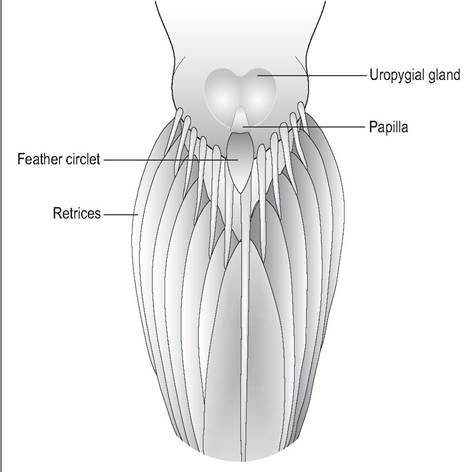
Figure 6.72 • Dorsal view of uropygial gland. Surrounding feathers have been plucked for better visibility.
patch loses feathers under the influence of estrogen and becomes thickened and vascular to provide extra warmth during egg incubation. In some species like gulls the number of brood patches is matched to the number of eggs in the clutch.