Intervertebral Disks
Intervertebral disks are present in every intervertebral space except that between the first and second cervical vertebrae (p. 36). These disks are functionally important because of their contribution to the flexibility of the spine and to the distribution of pressure over the extremities of the vertebrae.
Some degenerative changes occur normally with age, including the metaplastic changes of fibrous tissue, the calcification of the gelatinous nucleus, and, frequently, the separation of the fibrous lamellae of the anulus, the narrow dorsal part of the anulus being most vulnerable. When degeneration is severe, as occurs in particular dog breeds (see later), stretching or total rupture of the dorsal part of the annulus allows disk material to protrude into the vertebral canal, where it may put pressure (through the meninges) onto the spinal cord and nerves, resulting in various and often severe neurologic dysfunctions.The dorsal longitudinal ligament is well developed in the cervical region, preventing dorsal herniation of disk material into the vertebral canal. In this region, the degenerating disk material protrudes dorsolaterally toward the roots of the spinal nerves, resulting in their compression. Approximately 15% of disk problems in dogs occur in the cervical region; the clinical signs are neck pain, spasms of shoulder muscles, and lameness due to pain referred to a forelimb. The presence of the intercapital ligaments (between the heads of a rib pair passing beneath the dorsal ligament) at the joints T1-T2 to T9-T10 offers almost complete protection against herniation to the greater part of the thoracic cord (see Fig. 2.18). Thoracolumbar lesions account for the remaining 85% of intervertebral disk problems (T11/12 to L1-L2). In the caudal thoracic and lumbar regions of the spine, where the dorsal longitudinal ligament is thinner, dorsal protrusions and consequent spinal cord compressions are more frequent.
Common radiographic findings in cases of disk herniation are narrowing or collapse of the intervertebral disk space, collapse of the synovial joints, narrowing of the intervertebral foramen, and calcified material within the vertebral canal.
Misinterpretation of apparent narrowing of intervertebral disk spaces is easy if insufficient attention is paid to the geometry of image formation (p. 5). Furthermore, it should be emphasized that nuclear calcifications are often evident in radiographs of dogs that cause no signs of dysfunction or pain. The intervertebral disks of cats are not immune to degeneration, but for reasons that are obscure, affected animals very often fail to manifest any clinical signs.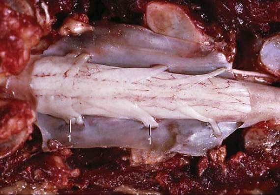
FIG. 12.8 Dorsal view of opened vertebral canal (cat). 1, Spinal nerves penetrating arachnoid and dura mater.
There are both breed and regional differences in the incidence of disk pathology. Chondrodystrophic breeds, such as the Dachshund and Pekingese, in which the degenerative process is both precocious and accentuated, are particularly prone to protrusions at a relatively early age. In normal dogs, disk disease is characterized by slow fibroid degeneration, most evident between 8 and 10 years of age; mineralization of the disk is unusual. As a result of chronic degenerative disk disease (without clinical signs), spondylosis may develop. The sites most frequently involved are those undergoing the greatest mechanical stress. As a result of the stress, bony spurs are formed ventral and lateral to the intervertebral disk space, leading ultimately to complete fusion of vertebrae. On survey radiographs the presence of spondylosis is often regarded as an incidental finding.