LUMBAR VERTEBRAE
The lumbar vertebrae continue several features of the thoracic vertebrae. Their bodies are about twice as long as those of the first thoracic vertebrae and are characterized by long transverse processes that sweep cranio- ventrally, overlapping the preceding vertebra (Figure 12-7/1).
The ventral deflection of these processes is even more pronounced in the cat. The interarcuate spaces of both lumbar and thoracic segments are very small, which makes access with a needle to this part of the vertebral canal difficult. The space at the lumbosacral junction is much better suited for this purpose. It is about 1 cm in diameter (in medium-sized dogs) and lies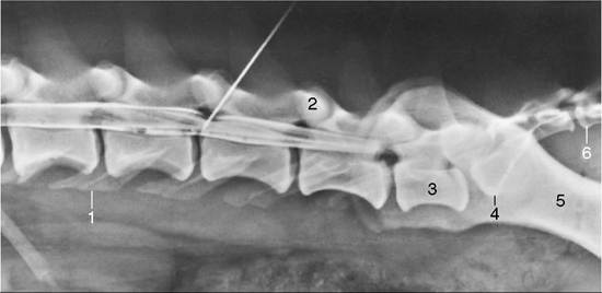
Figure 12-6 Lateral radiograph of the lumbar area of a dog with a myelogram. A needle is in the interarcual space between L4 and L5. 1, Transverse processes of L4; 2, articular processes; 3, last lumbar vertebra (L7); 4, promontory (of sacrum); 5, shaft of ilium; 6, first tail vertebra (Cdl).
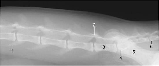
Figure 12-7 Lateral radiograph of the lumbar region of a cat. 1, Transverse processes of L4; 2, articular processes; 3, last lumbar vertebra (L7); 4, promontory (of sacrum); 5, shaft of ilium; 6, first tail vertebra (Cdl).
in the transverse plane of the highest palpable points on the wings of the ilia but about 2 cm deeper. In the cat, the interarcuate space between the last two lumbar vertebrae is also wide enough to allow injection into the vertebral canal.
The mamillary processes are also fused with the cranial articular ones in the lumbar regions.