THIRD TO SEVENTH CERVICAL VERTEBRAE
The spinous processes of the remaining cervical vertebrae increase in height and in cranial inclination. The ventral crests are most prominent at the caudal ends of the bodies, marking the positions of the intervertebral disks directly caudal to them.
The transverse processes have distinct cranial and caudal extensions (ventral and dorsal tubercles). The ventral tubercle of the sixth ver-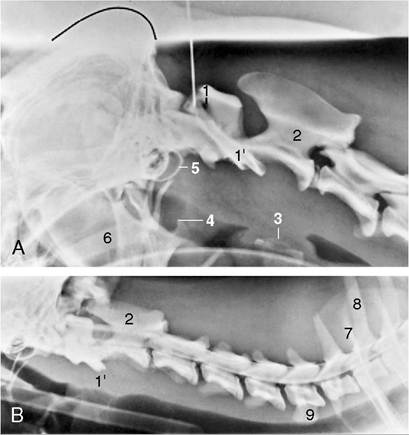
Figure 12-3 A, Lateral radiograph of the head-neck junction of an intubated dog. Note the needle in the atlantooc- cipital space for a cerebrospinal fluid tap. The dorsal contour of the skull is marked. B, Myelogram of an intubated dog. 1, Lateral vertebral foramen of atlas; 1', wing of atlas; 2, axis; 3, cricoid cartilage; 4, angular process of mandible; 5, tympanic bulla; 6, soft palate; 7, spine of scapula; 8, spinous process of T1; 9, ventral tubercle of C6.
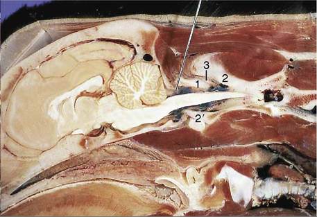
Figure 12-4 Median section of head and neck (dog); the needle penetrates the atlantooccipital membrane to enter the subarachnoid cerebellomedullary cistern. 1, Dorsal arch of atlas; 2, spinous process of axis; 2', dens; 3, dorsal atlantoaxial ligament.
tebra is a nearly sagittal plate that projects considerably below the contour of the body (Figure 12-3/9). The transverse process of the seventh is a rodlike lateral projection that does not overlap the body ventrally. The caudal extremity of that body bears an articular fovea for the head of the first rib. The flat articular surfaces of the synovial joints are nearly horizontal. The cranial
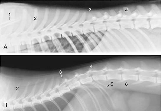
Figure 12-5 Lateral radiographs of canine (A) and feline (B) thoracic and lumbar vertebrae.
Radiograph A was obtained after the injection of a contrast agent into the subarachnoid space. 1, Scapular spines; 2, spinous process of T5; 3, anticlinal vertebra (T11); 4, spinous process of L1; 5, rudimentary rib; 6, sublumbar muscles.articular processes, which provide the ventral component of these joints, narrow the large intervertebral foramina from above.
The wide joint spaces of the atlantooccipital and the atlantoaxial joint support relatively free vertical and rotational movements. The nuchal ligament extends from the spinous process of the axis to the tip of the first thoracic spinous process; it is then continued by the supraspinous ligament until the third sacral vertebra. The nuchal ligament plays an important role in the support of the head of the dog and must be spared during surgery (see Figure 2-8/5). The ligament is not present in cats, but they do possess a supraspinous ligament.
Disorders of the cervical vertebral column, producing compression of the spinal cord, occur in large dogs, especially the Great Dane and the Doberman Pinscher. These disorders may involve deformation of the vertebral arch, malformation of articular facets, vertebral instability of C5-C6 or C6-C7, and dorsal displacement of the vertebral body.
Thoracic vertebrae
The bodies of the thoracic vertebrae are relatively short but increase in length from the tenth caudally (Figure 12-5). The long spinous processes of the first half of the thoracic region are of about equal length. Those of the second half gradually decrease in height; their caudal inclination changes at the eleventh thoracic, the anticlinal vertebra. A more noteworthy change occurs in the orientation of the articular surfaces. On the first 10 (or so) thoracic vertebrae these surfaces lie roughly in a dorsal plane (like those of the cervical vertebrae); caudal to this they are nearly sagittal, and the cranial articular processes enclose the caudal ones (see Figure 2-10). The articular spaces of the former joints are best depicted in lateral radiographs (Figure 12-6), and those of the latter, in ventrodorsal radiographs. The more cranial thoracic vertebrae favor lateral movement of the column, whereas the more caudal bones favor sagittal flexion and extension. Other features of the canine and feline vertebrae are the presence of the mamillary and accessory processes. The mamillary processes are short dorsal projections of the transverse processes that first appear at the third thoracic vertebra and, from the eleventh, migrate dorsally to surmount the cranial articular processes. The accessory processes arise from the caudal border of the pedicle and are present from the midtho- racic to midlumbar regions; they are confined to the last three thoracic vertebrae in cats (see Figure 2-11/1,2).