LYMPHATIC STRUCTURES
The lymphatic system has two components. The first comprises a system of lymphatic capillaries and larger vessels that return interstitial fluid to the bloodstream. The second comprises a variety of widely scattered aggregations of lymphoid tissue, including the many lymph nodes; less discrete lymphoid aggregations, such as tonsils, are not considered until later (p.
257).Lymphatic Vessels
A plexus of lymphatic capillaries that is spread through most tissues collects a fraction of the interstitial fluid. This fraction is disproportionately important because it includes the proteins and other large molecules that are unable to enter the less permeable blood vessels. The greater permeability of the lymphatic capillaries also allows them to take in particulate matter, including microorganisms on occasion. The lymphatic capillaries commence blindly and form plexuses from which larger lymphatic vessels take origin. These larger vessels closely resemble veins in structure but are more delicate. Because the fluid (lymph) they contain is generally pale, they are rarely conspicuous; however, they are easily identified once seen, as closely spaced valves give them a distinctive beaded appearance when they are well filled. The largest vessels take independent courses, but many of smaller size accompany blood vessels and nerves. The lymphatic vascular tree eventually converges on two or three large trunks that open in a rather erratic fashion into major veins at the junction of the neck and thorax (Figure 1-34).
Lymph Nodes
Lymph nodes, often incorrectly termed lymph glands, are placed along the lymph pathways in a pattern that shows considerable specific and some individual variation. Groups of neighboring nodes constitute lympho- centers, whose occurrence and drainage territories exhibit greater constancy than is presented by individual nodes.
There are important interspecific differences in the lymphocenters: those of the domestic carnivores and ruminants each contain rather few but individually large nodes, particularly in cattle, and those of pigs and, more especially, horses each contain a great many small nodes packeted together.Lymph nodes are firm, smooth-surfaced, and generally ovoid or bean-shaped. Some that are superficial can be identified on palpation through the skin. Naturally, they are more easily found when they are enlarged, and it is therefore a matter of importance to have a clear expectation of which nodes can usually be identified in the healthy animal. Each node is bounded by a capsule, below which runs an open space (subcapsular sinus) into which the afferent vessels open at scattered sites. Branches from the subcapsular sinus lead to a medullary sinus close to the generally indented hilus, where the few efferent vessels emerge (Figure 1-35, A; see also Figure 7-50). The tissue of the node is divided between cortical and medullary regions. The cortex contains the germinal centers in which lymphocytes are continually produced; the medulla consists of looser branching cellular cords. Both are supported by a reticular framework containing many phagocytic cells. The organization of the lymph nodes of pigs (Figure 1-35, B) shows a reversal of the usual flow pattern: the afferent vessels enter together, whereas the efferent vessels have dispersed origins (Figure 7-51, A-B).
With very few exceptions (and these are disputed), all lymph passes through at least one node in its passage from the tissues to the bloodstream. As it percolates through the node, it receives a recruitment of lymphocytes and is also exposed to the activities of the phagocytes. These remove and destroy, or attempt to remove and destroy, particulate matter, including any microorganisms within the lymph. The lymph node thus provides a barrier to the spread of infection and tumors, some varieties of which favor lymphatic pathways for their dissemination.
Swelling of a lymph node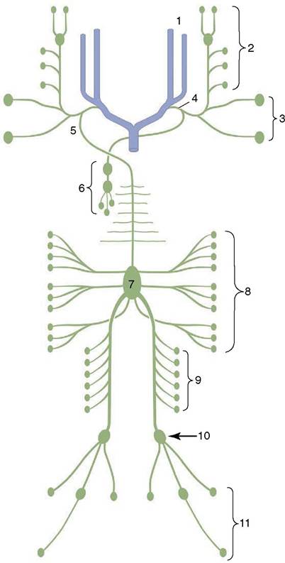
Figure 1-34 Generalized schema of the lymph nodes and lymphatic vessels (dorsal view). The top of the diagram represents the neck region. 1, External and internal jugular veins; 2, lymph from the head; 3, lymph from the shoulder and forelimb; 4, tracheal duct; 5, thoracic duct; 6, lymph from the thoracic organs; 7, cisterna chyli; 8, lymph from the abdominal organs; 9, lymph from the lumbar region and kidneys; 10, lymph nodes of the pelvis; 11, lymph from the hindlimb.
frequently indicates the existence of a disease process in its drainage territory. It is clear that the role of the lymphatic system in disease is equivocal. On the one hand, lymph flow facilitates the spread of microorganisms or tumor cells; on the other, the intervention of the node provides an opportunity for their containment and destruction. There are obviously weighty reasons why the position, the accessibility, the drainage territory, and the destination of the efferent flow of all major nodes must be familiar to the clinician,
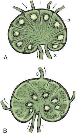
Figure 1-35 Structure of a lymph node (A) in which the germinal centers (lymph nodules) occupy the cortical region. In the pig (B) the germinal centers lie centrally. The arrows indicate the direction of lymph flow. 1, Afferent lymphatics; 2, subcapsular sinus; 3, efferent lymphatics.
the pathologist, and the veterinarian engaged in meat inspection.