PERIPHERAL BLOOD VESSELS
The peripheral blood vessels comprise arteries that lead blood from the heart, veins that return blood to the heart, and capillaries that are the minute connections between the smallest arteries and the smallest veins within the tissues.
These vessels are arranged to form two circuits (Figure 1-31). One, the greater or systemic circulation, arises from the left ventricle, conveys oxygenated (arterial) blood to all organs and parts of the body other than the exchange tissue of the lungs, and then transports the now deoxygenated (venous) blood back to the right atrium; the second, the lesser or pulmonary circulation, conveys deoxygenated blood from the right ventricle to the exchange tissue of the lungs, where it is reoxygenated before being returned to the left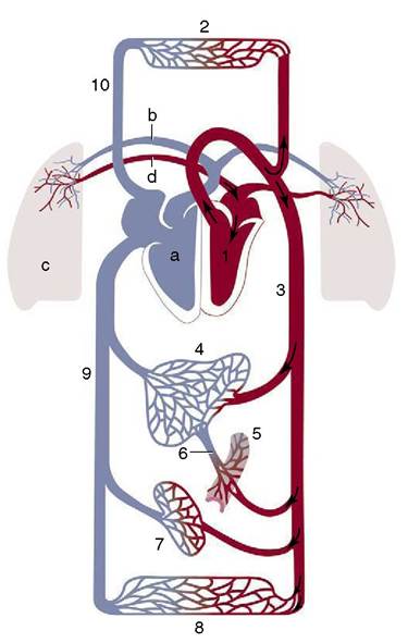
Figure 1-31 Schema of the circulation; vessels carrying oxygenated blood are shown in red, those carrying deoxygenated blood in blue. Systemic circulation: 1, Left side of the heart; 2, vessels in the cranial part of the body; 3, aorta; 4, liver; 5, intestines; 6, portal vein; 7, kidneys; 8, vessels in the caudal part of the body; 9, caudal vena cava; 10, cranial vena cava. Pulmonary circulation: a, Right side of the heart; b, pulmonary artery; c, lung; d, pulmonary vein.
atrium by a special set of veins. The systemic and pulmonary circulations together with the chambers of the heart form a single complex course through which the blood circulates endlessly.
Arteries
In the dissection room, the arteries may be distinguished from other vessels by their white, thick, and relatively rigid walls and their empty lumina (unless filled with an injection mass for the convenience of the dissector). The larger arteries follow a rather constant pattern, but their smaller branches show much variation—so much so that some patterns described in the textbooks, though the most common, may actually occur in only a minority of subjects.
When arteries branch, the combined cross-sectional area of the daughter vessels always exceeds the cross section of the parent trunk (Figure 1-32).A general correspondence exists between the absolute and relative sizes of parent and daughter vessels
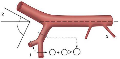
Figure 1-32 The branching of the arteries. Note that (1)the sum of the cross-sectional areas of the branches always exceeds that of the parent trunk; (2) large branches leave the trunk at more acute angles than smaller branches; and (3) the smallest branches leave erratically.
and the angles at which the latter diverge from the main trunk. Although there are exceptions, larger branches diverge at more acute angles to minimize resistance. Hemodynamic factors are less important where small branches are concerned, and these often follow the shortest routes to their destinations (see Figure 1-32).
Another factor influencing arterial course is a preference for protected situations; this is well illustrated in the limbs, where the major vessels tend to run medially and also tend to reorient themselves to cross the flexor aspects of successive joints. In comparable fashion, arteries that supply organs that change much in size or position are protected against stretching by taking meandering courses.
Although arteries ultimately discharge into capillary beds, most also have more proximal and more substantial connections with their neighbors. These interarte- rial connections (anastomoses) provide alternative, collateral pathways or bypasses by which circulation can be maintained when the more direct route is blocked. Collateral circulation operates as soon as a main trunk is obstructed and becomes more efficient with the passage of time.
The possibility for collateral circulation in different regions and organs has obvious importance to the clinician and the pathologist, and more attention is given to this topic later (p.
242). Meanwhile, this possibility suggests that it may be unnecessary to know the details of all the smaller vessels.Veins
In the dissection room, veins are distinguished by their thinner walls, their frequently collapsed appearance, and their capacity, which is invariably greater than that of the associated arteries. They appear blue when filled with clotted blood. Most veins are also distinguished by the presence of valves, which are repeated at intervals along their length; the valves ensure a unidirectional
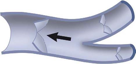
Figure 1-33 A branching vein opened to expose valves. The arrow indicates the direction of blood flow.
flow and prevent reflux of blood when the circulation stagnates (Figure 1-33). Each valve consists of two or three semilunar cusps facing each other. Valves are most numerous in veins that are exposed to intermittent changes in external pressure and are wholly lacking in those isolated from such influences. They are thus common in veins running between muscles and absent from those in the vertebral canal and cranial cavity; partly on this account, the veins in the latter site are known by the special term venous sinuses.
The very largest arteries and veins run separately, but most veins of medium and lesser size accompany the corresponding arteries to which they are said to be satellite. However, they show even more variation than do the arteries and are quite commonly duplicated, further replicated, or arranged in plexus formation.