Magnetic Resonance Imaging (MRI)
MRI scanning is a fairly recent development in veterinary practice, and it continues to make advances as the techniques are refined and applied. It is tempting to consider that MRI will render radiography out-dated, but this is a mistake as the techniques are
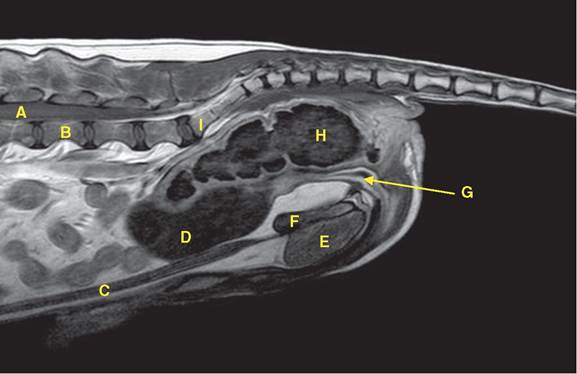
Figure 18.5 Lateral MRI scan of the pelvis of a dog.
A = spinal cord; B = 5th lumbar vertebra;C = penile urethra; D = urinary bladder; E = gracilis muscle; F = pelvic symphysis; G = urethra (male); H = rectum; I = sacrum.
complementary. Even computed tomography (CT), which is closely related to radiography, will not supersede radiography in the near future.
MRI scanning requires that the production of the images takes time and that the patient must remain quite immobile whilst the scanner builds the picture, taking 5-15 minutes. Of course, radiography takes a picture like a camera and the image is then developed without involvement of the patient. Heavy sedation or anaesthesia is required as it takes the scanner 5-30 minutes to construct the image.
The MRI scanner uses a strong magnetic field and radio waves to construct an image. The magnetic field is either from a permanent magnet or a superconducting electromagnet. The former is less expensive to buy and maintain but is not as powerful in the quality of results. It is not within the scope of this book to explain how the MRI scanner produces images (see the bibliography). However, it should be pointed out that permanent magnet systems operate at a low field strength, typically around 0.25 Tesla, whereas superconducting magnets, as used in medical and veterinary diagnosis, have a field strength of 0.5-1.5 Tesla (and even 3.0 T). Basically it is the relaxation of the excited hydrogen atoms in water and fat that changes the radio signal that is being induced. It is the recovery and direction of the spinning hydrogen atoms that results in T1 and T2 relaxation, the basis of the two main types of MR image. In T1 images water has a low (dark) signal and fat has a high (bright) signal: in T2 images water has a high (bright) signal and fat also has a high (bright) signal. Since the abdomen has high concentrations of both water and fat, it is not surprising that this region gives excellent opportunities for imaging.
MRI scanning is particularly helpful for locating and assessing neoplasia and foreign bodies (Figures 18.6 and 18.7). Sometimes it is helpful to administer a contrast agent with a T1 weighted sequence so that, for example, an abnormally vascular organ or tissue will have a bright image. Gadolinium is a contrast agent that is used in MRI scanning of veterinary patients, although it is not licensed for veterinary use. Figures 18.5-18.7 show examples of MRI scans of the abdomen and pelvis of canine patients.
18.4