Specific Organs
18.2.1 The stomach
The dark area of the stomach represents the gas-filled area of the fundus and is situated to the left of the midline at the level of the last two ribs. The distended stomach extends caudally to the ribs and can occupy almost a third of the abdominal cavity when distended.
Often the stomach has a figure-of-eight shape on lateral radiographs, indicating a state of contraction (Figure 18.2). When the stomach is not distended it is largely overlain by the liver (Figures 18.1 and 18.2).Oral barium is often administered to aid the diagnosis of tumours, diaphragmatic hernias or radio-transparent foreign bodies such as plastic toys or fur balls in cats. The radiograph should be obtained no longer than 3 minutes after administration of the barium when the stomach is being investigated. (See Section 18.2.4 for speed of transit of barium in the intestines.)
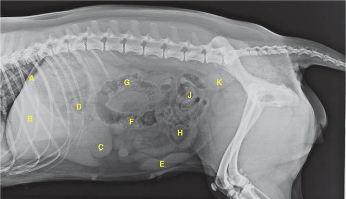
Figure 18.1 Lateral radiograph of the abdomen of a dog. A = diaphragm; B = liver; C = stomach; D = stomach; E = spleen; F = descending colon; G = ascending colon; H = urinary bladder;
J = jejunoileum; K = rectum.
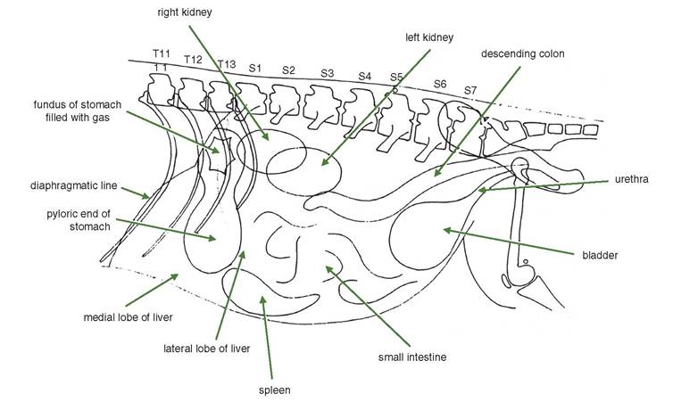
Figure 18.2 Diagram of lateral radiograph of the abdomen of the dog in the standing position. Rarely do all of the organs show as clearly as this. Alteration of the exposure values and times improves the visualisation of the different structures.
18.2.2 The spleen
The spleen does vary in position depending on the fullness of the stomach. It looks conspicuously dense compared with adjacent structures due to its high content of blood. The size of the spleen can vary markedly between normal individuals and can be considerably enlarged as a result of barbiturate anaesthesia.
18.2.3 The liver
The liver is easily recognised as an area of relative density, although its precise outline is readily determined. Its cranial boundary coincides fairly closely with the concave diaphragmatic line. The borders of the lobes tend to merge with the stomach and the spleen.
Cholecystography is a radiological procedure involving the administration of an iodine compound that is administered intravenously or orally and is preferentially excreted by the liver. The result is outlining of the gall bladder and the bile, hepatic and cystic ducts radiographically. Portal venography is a contrast technique used to facilitate the radiographic appearance of portosystemic shunts (see Section 7.7).
18.2.4 The intestines
The large intestine is often clearly visible radiographically due to the presence within its lumen of gas, ingesta or faecal matter showing as areas of varying radiotransparency or opacity. The small intestine is not so easily distinguished, but its presence is suggested by an area of mottled opacity and the occasional gas bubble.
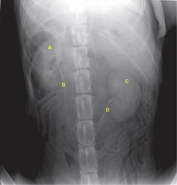
Figure 18.3 Ventrodorsal radiograph of the abdomen of a dog. A water-soluble contrast medium has been given intravenously to show the kidneys and ureters. A = right kidney; B = right ureter; C = left kidney; D = left ureter.
Oral barium is used to show the position, and often the cause of obstructions, of the small and large intestines. It is also used for detecting tumours and the displacement of the alimentary tract.
The region of the intestine containing barium, as seen on the radiograph, depends on the time between administration and exposure. Some barium is seen in the duodenum after only 5 minutes, and the whole of the small intestine is visible after 30 minutes. The ascending and descending colon is filled by 90 minutes, and the barium remains within the descending colon and the rectum until defaecation.
A better technique for the visualisation of the large intestine is the administration of a barium enema since immediate and post-evacuation radiographs give an accurate clinical study; the caecum is particularly well seen by this procedure.18.2.2 The kidneys
To obtain optimum radiography of the kidneys, the alimentary tract should first be emptied as completely as possible. The kidneys are well demonstrated on a lateral film, although there is some overlap (see Figure 18.2). In ventrodorsal radiographs there is
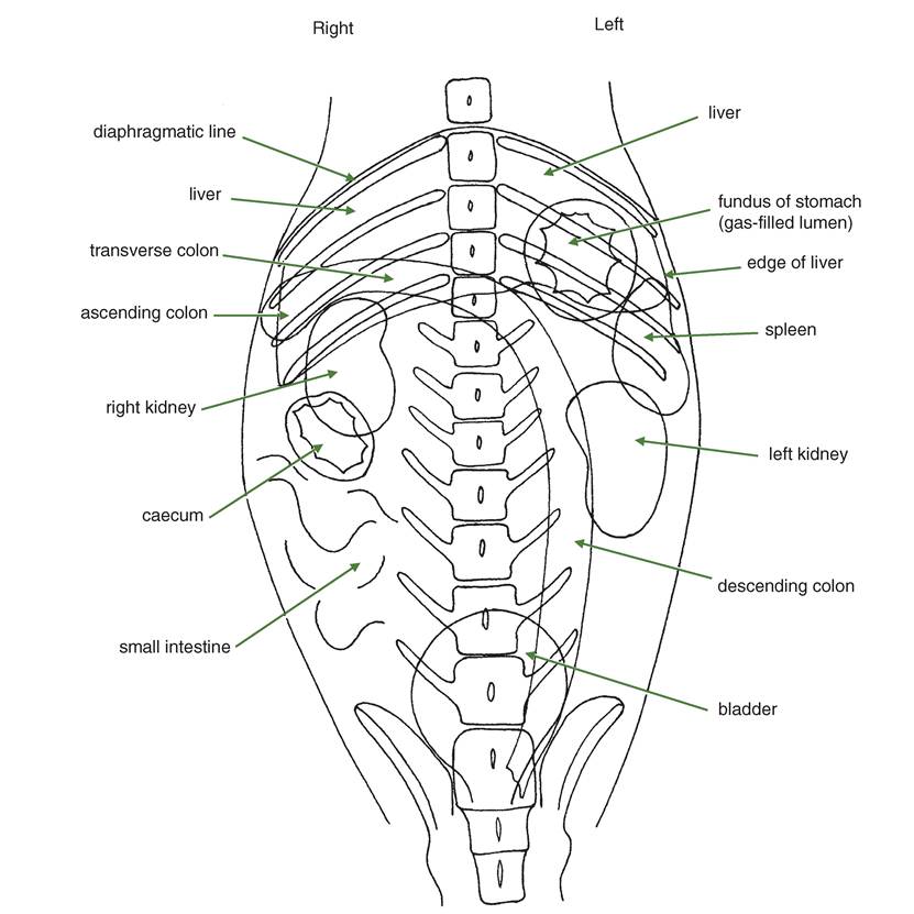
Figure 18.4 Diagram of ventrodorsal radiograph of the abdomen of the dog.
some obscuring superimposition by the liver (right), spleen (left), caecum (right) and the lumbar muscles. Variations in size, tumours and, sometimes, renal calculi may be identified without the use of contrast media.
Although there is little application in veterinary practice, the kidneys are well demonstrated by pyelography (urography). The technique involves the intravenous administration of an organic water-soluble iodide, preferably under anaesthesia. Apart from radiopacity of the kidneys, the renal end of ureters is rendered radiopaque.
The introduction of air into the peritoneal cavity does improve the external outline of the kidneys, but the patient does need to be in ventral recumbency.
18.2.3 Urinary bladder
The bladder is clearly visible in both lateral and ventrodorsal views, providing the bladder contains an appreciable amount of urine since it is difficult to see when empty.
Clinically radiography of the bladder is carried out most frequently for the purpose of detecting cystic calculi. However, urate stones, common in the Dalmatian, are not adequately radiopaque. Pneumocystography, a radiographic study following the introduction of air into the bladder via a urethral catheter, is a useful technique for detecting calculi.
Aqueous organic iodides may be introduced into the bladder by urinary catheter for the investigation of an obstruction or neoplasia.
18.2.4 Urethra and prostate gland
The urethra is not easily seen on plain radiographs, although its position is indicated by the os penis in male dogs. The main reason for its radiographic study is to demonstrate the presence of calculi, which are usually radiopaque. The prostate gland is not seen on radiographs unless it is markedly enlarged and it has dropped forward over the pelvic brim. In this position it must be distinguished from the urinary bladder.
18.3