Main genera and species of yeasts isolated from the oral cavity of dogs and clinical signs
Just like in humans, dogs have a known range of yeasts in their oral mucosa that still requires more studies regarding colonization and pathogenicity. Despite its remarkable importance in the health of dogs, studies involving the isolation and correct identification of yeasts began to be developed in the 20th century [26].
This microbiota is not yet fully described, due to its great complexity and diversity. Fungal colonization of the oral cavity of dogs is associated with yeasts of the genera Candida, Malassezia, Trichosporon and Rhodotorula. Less frequently, we can isolate yeasts of the genus Cryptococcus [27].
In a recent study conducted with 50 mixed breed dogs, a yeast profile was found, composed of Candida albicans (39.5%), C. parapsilosis (18.6%), C. zeyl- anoides (13.9%), C. krusei (7%), C. tropicalis (4.7%), Trichosporon spp. (4.7%), T asahii (4.7%), C. guilliermondii (2.3%), T mucoides (2.3%) and Malassezia pachydermatis (2.3%). The genus Candida showed a high prevalence, making up a total of 82.2% of the isolated yeast profile. It is worth mentioning here the isolation of Candida zeylanoides, a rare species, even in humans, and thus, the oral mucosa of dogs can harbor a new “ecological niche” of this fungus species, which can also act as an opportunistic pathogen [9].
5.1 Genus Candida and Candidiasis
Currently, 317 species of this genus are recognized. Several of these species, more precisely 20, have a pathogenic potential and can thus cause infections in several species of animals, such as dogs [17]. The relationship with the host can be commensal, parasitic or saprophytic. It can also be found in the usual form of a yeast, or in the form of pseudohyphae. Candida albicans is the most common colonizer in cases of infections, with a predilection for mucous surfaces and mucocutaneous areas. Other species, such as C.
kefyr, C. lusitaniae, C. guilliermondii, C. tropicalis, C. krusei, C. famata, C. parapsilosis, can be isolated from animals (Figure 5) [15].There were only few cases found in the literature in small animals, however, reports of candidiasis in various animal species are also increasingly common, described in photos of pyoderma of the lip folds, disseminated and localized mucocutaneous dermatitis, urinary tract infections, gastrointestinal and reproductive system, ear infections, systemic and oral infections [27].
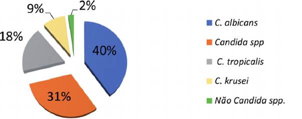
Figure 5.
Presumptive result of Candida species isolated from the oral cavity of mixed breed dogs according to the Chromagar Candida®.
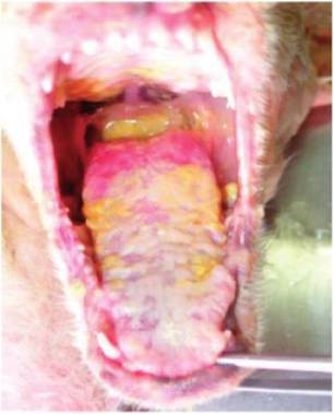
Figure 6.
Mixed breed dog with oral candidiasis (glossitis). Friable white and yellowish plates covering the tongue [28].
Candidiasis related to the digestive system of dogs, such as a clinical manifestation of glossitis, is characterized by the formation of pseudomembranous plaques, usually whitish in color, or yellowish beige. Once these plaques were removed, we noticed erythematous regions with the presence of ulcers (Figure 6) [28].
3.1 Genus Trichosporon and Trichosporonose
The genus Trichosporon has 37 species that inhabit different ecological niches, such as water, soil and body and mucous surfaces of humans and animals. They can cause superficial and deep infections, such as Trichosporon asahii, T mucoides, T ovoides, T inkin, T asteroides and T cutaneum [29].
No cases of Trichosporon infections have been reported in the oral mucosa of dogs, however, several species have already been isolated as colonizers. Clinical cases of nasal granuloma in other animals, cystitis in cats, mastitis in cows and dermatitis in horses and monkeys have already been described (Figure 7) [30].
3.2 Genus Malassezia and Malasseziosis
The genus Malassezia has 15 species, mostly lipophilic yeasts, that can be part of the skin and mucous membranes of humans and dogs.
They are opportunistic yeasts, and in certain circumstances, they can lead to clinical manifestations [31]. Malassezia pachydermatis, the most frequent in dogs, is not lipophilic and can grow in a culture medium common in mycology [19] (Figure 8).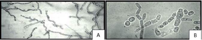
Figure 7.
A and B-Yeasts of Genus Trichosporon showing oval and rectangular artrospores - (A) 160x and (B) 400x [15].
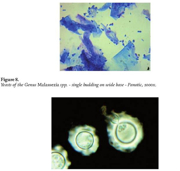
Figure 9.
Cryptococcus spp. Encapsulated yeasts - Nigrosina, 1000x.
Several clinical symptoms can be associated with Malassezia spp. and, particularly, in cases of otitis and dermatitis in dogs. Cases of otitis by Malassezia pachyder- matis are frequency, but oral infection caused by this agent have not been described or has not yet been well studied [19].
5.4 Genus Cryptococcus and cryptococcosis
In the Cryptococcus genus, we found 38 species, with Cryptococcus neoformans and C. gattii being the most prominent in medical mycology in man and animals (Figure 9). The species can be found in different places in the environment, primarily in association with birds’ droppings, mainly pigeons, but have an ecological association with trees too, such as eucalyptus [32].
In dogs, can enter the body through the lung causing pulmonary disease, and several clinical signs can be presented, such as skin lesions, nasal mucosa (“clown nose”), and can hit the central nervous system, for its neurotropic nature. These lesions in the nasal mucosa can extend into the oral cavity of the animals [28].
6.