Masticatory muscle function
Key point
■ The majority of the masticatory muscles are innervated by the motor nucleus of CN V and the mandibular branch of the trigeminal nerve. The caudal half of the digastricus muscle is innervated by cranial nerve VII.
The location of the motor nucleus of CN V is in the mid brainstem at the junction between the pons and the medulla oblongata (see Figs. A3, A30). Its efferent fibres travel only in the mandibular branch of the trigeminal nerve CN V and supply the masticatory muscles such as the temporalis, masseter, pterygoid muscles and mylohyoideus muscles that raise the mandible. It also supplies the tensor tympani muscle, used to protect the ear from loud noise, and the tensor veli palatini muscle which tenses the soft palate. The mandibular branch of CN V supplies the rostral belly of the digastricus muscle which opens the jaws, while the caudal belly is supplied by CN VII. The dual innervation of the digastricus muscle (CNN V and VII) reflects its embryonic origin from two pharyngeal arches. The facial nerve (CN VII) also supplies the buccinator muscle of the cheek, which functions to return food from the oral vestibule to the masticatory surface of the teeth.
The most common sign of damage to masticatory muscle innervation is atrophy (Fig. 10.12). This is due to LMN damage, for example, due to a tumour or neuritis. However, primary muscle disease, such as masticatory myositis, can also cause marked muscle atrophy. Lesions of CN V, mandibular branch, need to be bilateral to result in overt loss of jaw muscle tone and a ‘dropped jaw’.
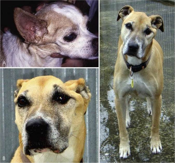
Fig. 10.12 Loss of LMN (peripheral motor unit) innervation to the masticatory muscles in these two dogs, resulted in severe atrophy of the temporalis and masseter muscles and prominence of the zygomatic arch. Both dogs had ipsilateral facial hypoalgesia. (C) Depicts the same dog as in (B), 2 months later
illustrating development of a head tilt. The dog also had nystagmus and ipsilateral facial paresis indicating involvement of CNN VII and CNVIII in the lesion. The dog in (A) had a tumour; the same was presumed for the dog in (B) and (C).