Sensory input from the head
Key points
■ All three branches of the trigeminal nerve transmit sensory information from the head to the sensory nucleus of CN V, which extends throughout the brainstem.
The trigeminal ganglion is located at the base of the neurocranium.■ Proprioception, tactile and thermal sensation, and nociception are received by the mesencephalic, pontine and myelencephalic regions of the sensory nucleus of CN V, respectively.
■ Tactile stimulation of the skin of the maxilla, the upper eyelid and the mandible tests the maxillary, ophthalmic and mandibular branches of the trigeminal nerve during the neurological examination.
Tactile, proprioceptive, thermal and nociceptive input from the head is received in the sensory nuclear complex of the trigeminal nerve in the brainstem (Fig. 10.2). All three branches of CN V (mandibular, maxillary and ophthalmic) are associated with sensory input from the face (Fig. 10.13). Sensory input from the external ear is via CN VII, IX and X. Lesions of CN V result in hypoalgesia or anaesthesia of the face. Sensory nerve cell bodies are located in the trigeminal ganglion outside the CNS; this arrangement is similar to sensory input via spinal nerves. Unusually, some somata are also sited in the CNS in the mesencephalic nucleus of V. The trigeminal (GasserianZsemilunar) ganglion is located in the trigeminal canal at the apex of the petrous temporal bone, inside the neurocranium.
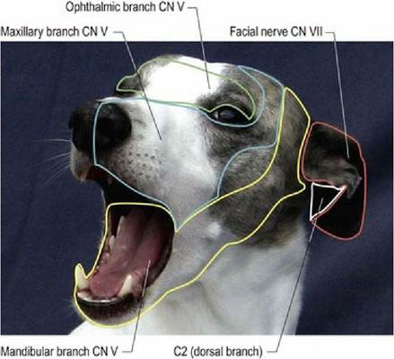
Fig. 10.13 The autonomous zones of facial sensory innervation. For the branches of the trigeminal nerve (CN V), green outlines the region for the ophthalmic branch, blue for the maxillary branch and yellow for the mandibular branch. The inner and outer aspects of the pinna are innervated by the facial nerve and branch of the second cervical nerve, respectively.
Sensory nucleus of CN V
The sensory nucleus of V complex is an elongated grey matter column that extends caudally from the midbrain to the substantia gelatinosa, which caps the dorsal horn of the spinal cord (see Fig. 6.6). The nuclear complex is associated with an adjacent white matter tract forming the spinal tract of V. The complex is divided into mesencephalic, pontine and spinal nuclei and tracts. It receives somatic afferent information (see Figs. A19-31).
Via CN V, the mesencephalic nucleus and tract of V receives proprioceptive input from the muscles of mastication, temporomandibular joint, teeth and, probably, the extraocular, facial and lingual muscles. Efferent axons travel to muscles of mastication and, via the reticular formation, to motor nuclei of the brainstem and cranial spinal cord.
The pontine nucleus and tract of V receives input about touch and pressure (mechanoreception). Collateral axons also project to motor nuclei associated with corneal, palpebral, tongue and salivary reflexes.
The spinal nucleus and tract of V is located, caudal to the attachment of CN V in the medulla oblongata where it is prominent in the dorsolateral area. It receives sensory input related to nociception and temperature from the skin and mucous membranes of the head. The main input comes in via CN V but some input from the ear is delivered via CNN VII, IX and X. The nucleus also contains interneurons connecting the trigeminal nucleus with other nuclei of the brain stem and cervical cord. The spinal nucleus and tract of V continue into the spinal cord as the substantia gelatinosa and dorsolateral fasciculus, which receive input from the body about noxious and thermal stimuli (see Table. 4.3).
For conscious perception of sensation from the head, axons from the sensory nuclear complex form the trigeminal lemniscus (quintatothalamic tract), decussate, join with the medial lemniscus and travel via the thalamus to the somatosensory cortex (see Fig. 6.1).
Lesions cause hypoalgesia, which is ipsilateral if the lesion is located in the brainstem, but contralateral if the lesion is located in the forebrain (see cases described in Fig. 10.12).