MHC Class Il Antigens on Antigen- Presenting Cells Play a Major Role in Selective Activation of T-Helper Cells
The expression of cell surface MHC class II antigens is highly restricted. They are only present on select types of cells includ-
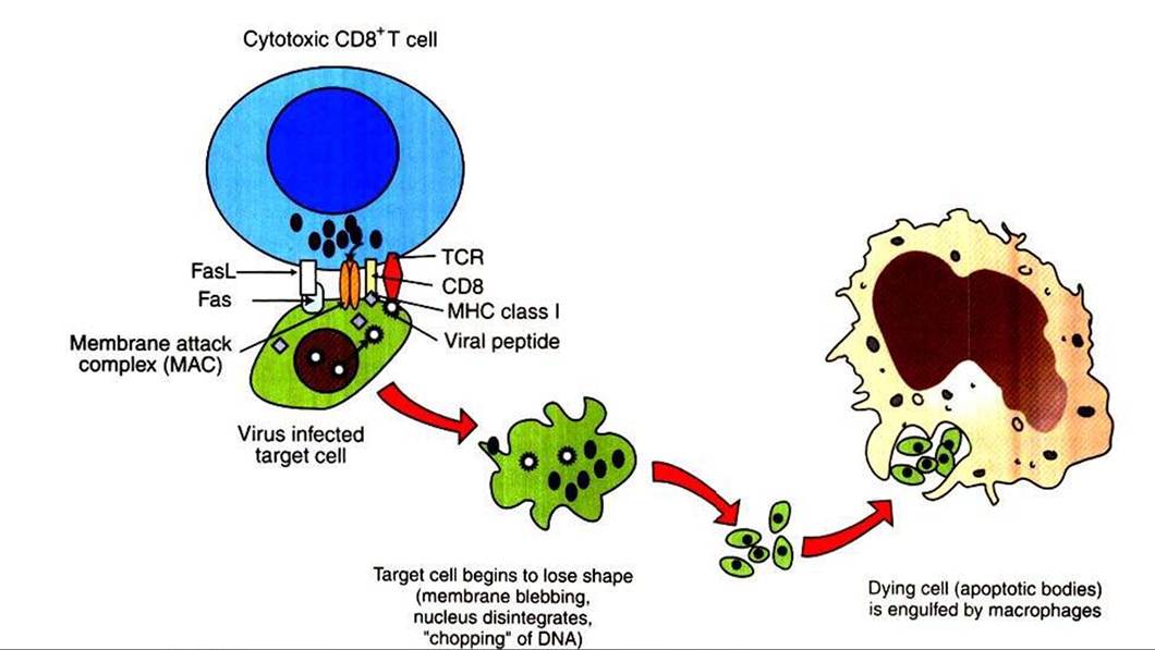
FIGURE 55-2 Mechanism of CD8-mediated cytotoxicity.
FasLf Fas ligand; MHCf major Nstocompatability complex; TCRf T-cell receptor.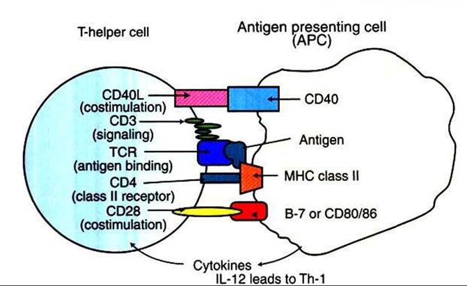
FIGURE 55-3 Interacting molecules of T-helper cells and antigen-presenting cells. IL, Interleukin; MHCt major histocompatibility complex; TCRt T-cell receptor; Th-IzTheIperceII type 1; Th-2, T-helper cell type 2.
ing dendritic cells, select macrophages, B cells, and keratino- cytes. The presence of class II antigens on their surfaces endows these cells with a unique ability to present antigens to CD4* Th cells (Figure 55-3). Therefore these cells are called professional APCs. Class II antigens are two-chain molecules composed of a glycoprotein, 33-kD α chain and a shorter, 27-kD β chain that form a groove onto which processed (exogenous) antigenic peptides bind. For example, when a macrophage phagocytoses an antigen and breaks it down into peptides within a vacuole, intracellular MHC class II antigens bind to these processed peptides, and this complex moves to the surface of the cell to be presented to CD4+ T cells. The processed antigenic peptide specifically binds to the TCR on T cells, and the class II MHC proteins (on APCs) specifically interact with the CD4 molecule on T cells. These interactions are the first steps in activation of Th cells. Activation of T cells is highly regulated because their inadvertent activation has profound and widespread consequences; the cytokines secreted by activated Th cells can affect a wide range of both lymphoid and nonlymphoid cells.
Activation of CD4+ T cells requires at least two signals for activation. The primary activation signal determines the specificity through interactions of antigenic peptides and MHC molecules on APCs with the TCR∕CD3 complex on T cells. The second signal is referred to as a Co-stimulatory signal. Costimulatory signals include interaction of CD28 and/or CD40L, both residing on T cells, with CD80∕86 and/or CD40, both residing on APCs. Cytokines that are released from APCs, such as IL-12, IL-18, and IL-27, promote the generation of a Th-1 subset of cells.
Activation of T cells is strictly controlled, with two major restrictions. First, T cells cannot recognize free antigens; rather, they recognize short peptides that are a product of processed antigens by the APCs. Second, the processed antigen must be physically associated with the MHC molecules. This results in molecular interactions of antigenic peptide bond to MHC class I or class II molecules on cells or APCs, with the TCR and CD8, or TCR and CD4 on T cells, respectively.
As mentioned, T cells will specifically interact with antigenic peptides through specific recognition by the TCR on T cells, analogous to the B-cell receptor (BCR) on B cells. The TCR on T cells belongs to the immunoglobulin superfamily and thus has variable and constant regions along with transmembrane and cytosolic domains. Similar to B cells, the variable portion of the TCR chain determines the specific binding to the antigenic peptide. Because more antigens exist in the universe than the actual number of T cells, these cells have adapted a variety of molecular mechanisms to interact specifically with an innumerable number of antigens. These mechanisms include recombination of TCR genes (similar to the BCR), unequal sister-chromatid exchange, and nucleotide insertion at various locations of the variable gene segments. An important difference between the TCR and BCR is that the TCR does not undergo somatic mutation. If the TCR could undergo somatic mutation, there would be an increased chance of inadvertently generating a TCR reactive against “self” antigens, resulting in devastating autoimmune conditions. This attribute is critical for survival because T cells, unlike B cells, can affect a large number of diverse lymphoid and nonlymphoid cells through secretion of cytokines.