Microscopic Study of the Cell
Cells range in diameter from about 10 to 100 μm (micrometers). Cells that are actively multiplying range from about 20 to 30 μm in diameter. Table 2-1 lists the relationships among metric units of measurement used for microscopy.
(For example, 1 μm is one-thousandth of a millimeter, and there are about 25 mm in 1 inch; thus, approximately 1000 cells, each 25 μm in diameter, could be lined up between the 1- and 2-inch marks of a ruler.) sizes of cells vary considerably from one type of cell to another, but with the exception of the yolks of birds’ eggs (which are considered single cells), the distance from the center of the cell to some portion of the cell membrane (surface of the cell) is seldom more than a few micrometers.The outer cell membrane is thin, 7-10 nm. Regardless of its composition, a membrane of this dimension can have little tensile strength; this is another reason cells must be small.
The uniformly small size of cells and the much smaller sizes of structures within the cell have made effective study of cells difficult. As noted earlier, the existence of cells was not confirmed before the microscope was invented.
Details of the actual structure of the various parts of cells were not known with any degree of certainty until after the development of the electron microscope. The study of gross anatomy goes back several centuries, but understanding of the finer structure of the animal body awaited more recent technological developments.
Light Microscopy
some cells are in tissues that are thin enough to be illuminated from one side and observed with a microscope from the opposite side. This is true of the web of the foot of the frog, the mesentery of the intestine, and a few other tissues. in these instances, living cells can be observed directly, and this technique is useful for the study of blood circulation.
specific cells may also be taken from a living animal and grown on artificial medium by tissue culture. These cells may then be studied in the living state, even at high magnifications.Except for the forgoing situations, cells usually are studied after undergoing some degree of manipulation, so that what is actually seen with the microscope bears little resemblance to the living cell. A typical treatment of tissue before it can be examined with a light microscope includes the following:
1. Fixation with an agent, such as an aldehyde, that will cross-link the tissue proteins and prevent further changes in the tissue, such as autolysis and bacterial degradation. Alternatively, tissue may be frozen in liquid nitrogen to prevent such degenerative changes.
Table 2-1. Metric Linear Measurements
2. Embedding the tissue in a material that will permit cutting very thin sections. Paraffin is used for producing sections of 5-10 μm thickness; sections as thin as 12 μm can be obtained by embedding in a plastic, such as glycol methacrylate. Since most embedding media are not water soluble, the fixed tissue must be dehydrated and then infiltrated with some material such as xylene, which is miscible with the embedding medium. Frozen tissues need not be embedded.
3. Sectioning the tissue into very thin slices so that the sections may be placed on a glass slide. A microtome is used for this purpose. it consists of a sharp blade and a mechanism for moving the tissue past the blade and then advancing it a defined distance after each cutting. Frozen tissues are sectioned in a cryostat, which is a microtome housed in a freezer cabinet.
4. Staining the section so that different cells or different parts of cells can be differentiated according to color. Hematoxylin and eosin are stains commonly used together, and this treatment is described as an H & E stain. The hematoxylin tends to stain acidic portions of a cell dark blue or purple (these basophilic areas include the cell nucleus, which contains nucleic acids), and the eosin tends to stain the basic portions of a cell pink to red (these acidophilic areas include much of the more basic protein within the cell).
Wright’s stain, used to stain blood cells (Plates I and II), stains basophilic areas blue with methylene blue and acidophilic areas red with eosin. sections can also be treated with a variety of chemical solutions to demonstrate the presence of certain types of chemicals or the activity of enzymes in the tissue or cell, a technique called histochemistry (Fig. 2-6). The presence of specific types of molecules can be determined by exposing the section to a solution containing antibodies to those molecules. This technique is called immunocytochemistry.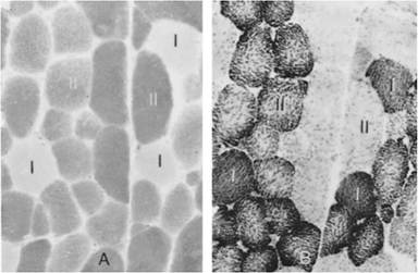
Figure 2-6. Serial sections of horse triceps brachii muscle histochemically stained for enzymatic activity. The same muscle cells (I and II) are visible in both sections. A) Calcium-dependent ATPase activity. B) Activity of a mitochondrial oxidative enzyme.
5. The last step, of course, is the actual examination of the stained section of tissue on the slide by means of a microscope and light transmitted through the section.
This approach to the study of the animal body has been standard for many years and will continue to be useful regardless of newer developments. However, some factors should be kept in mind when studying sections or photographs of sections.
The relationship of the tissue sections to the actual tissue is about the same as that of a sack of potato chips to a growing potato. Both the sections and the potato chips have been processed so that actual resemblance to the original structure is limited. Both are seen in two dimensions, length and width, with thickness relatively unimportant for visualization. Recent technological advances such as confocal microscopy and computer reconstruction provide three-dimensional views of cellular and tissue structure.
The light microscope can magnify objects to a maximum of about 1500 times the original size. This is known as the magnification, or power, of the microscope.
Resolving power refers to the property of showing two objects as separate structures. The light microscope can resolve (separate) two structures that are as close as approximately 0.2 μm (about 200 nm). This resolving power depends greatly on the wavelength of the light used to observe the tissue and the optical quality of the objective lens of the microscope.other developments in light microscopy include phase contrast and fluorescence microscopy. Phase contrast microscopy can be used with unstained and/or living cells, because it depends on differences in refraction of various parts of a cell for image formation. Fluorescence microscopy is often teamed with immunocytochemistry, in which antibody- labeled cells are identified by causing them to fluoresce upon exposure to light of a specific wavelength.
X-ray diffraction is used to study the structure of inorganic and organic crystals and the molecular structure of biologic substances such as DNA, collagen, and hemoglobin. it consists of passing a beam of x-rays through the substance and recording the diffraction pattern (scattering of the beam) on a photographic emulsion.
Electron Microscopy
Electron microscopes do not use visible light for the delineation of structures as in the light microscope; they use a beam of electrons focused by electromagnetic lenses. The electron beam may pass through a thin specimen in the transmission electron microscope or be reflected from the surface of an object and studied with the scanning electron microscope. The images with the electron microscope, however, are only black and white. (For an example of a scanning electron micrograph, see Fig. 15-2.)
The scanning electron microscope is a versatile instrument with a magnification range from ?15 to ?10,000 and a resolution in the vicinity of 10 nm. Depth of field with the scanning electron microscope is much greater than with any light microscope. Preparation of specimens for observation with the scanning electron microscope is relatively simple.
Non- metallic biologic material generally is dehydrated and coated with a thin layer of metallic gold before it is placed in the scanning electron microscope.The transmission electron microscope is capable of much higher magnification (as much as ?1 million), with an effective resolution of 0.1 nm. By the use of photographic enlargement and projection techniques, the magnifications can exceed 1 million and still show good detail. (A typical transmission electron micrograph is shown in Fig. 2-9.) Because so much more detail can be seen in a small area, tissue preparation for transmission electron microscopy is much more exacting and time-consuming than for light microscopy.
The best means of fixation is to apply a fixative (commonly glutaraldehyde followed by osmium tetroxide) to the living tissue or biopsy specimen. The time from the living state to immersion in the fixative should not exceed 2 minutes, and the size of tissue should not exceed 1 mm on a side. osmium tetroxide acts both as a fixative and as a stain. other heavy metals, including lead, may be used as so-called stains. The term stain may be used somewhat loosely, because the areas where the metals concentrate inhibit the passage of electrons, giving an electron-dense appearance that shows up as a dark area in the final photographic print. After fixation, the tissue is dehydrated and infiltrated with plastic and embedded in plastic for sectioning. The sections are cut extremely thin (less than 30 nm), placed on a grid, and examined with the electron microscope.
The picture of the typical cell viewed with the electron microscope still shows most of the structures described by light microscopy but in much greater detail. The typical cell seen in light microscopy consists of a nucleus and cytoplasm surrounded by the cell membrane (Fig. 2-7). The nucleus contains a nucleolus and chromatin material, which forms into chromosomes during cell division. A membrane called the nuclear envelope surrounds the nucleus. The cytoplasm contains a number of structures, or organelles, including the endoplasmic reticulum, Golgi apparatus, mitochondria, and inclusions.
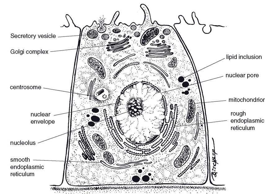
Figure 2-7. The general organization of a cell. The nucleus contains a distinct nucleolus. (Reprinted with permission of Wiley-Blackwell from Dellmann, H.D., Eurell, J. Textbook of Veterinary Histology. 5th edition. Baltimore: Lippincott, Williams & Wilkins, 1998.)