MICROSTRUCTURE OF SKELETAL MUSCLE
1. Is a muscle fiber the same as a muscle cell?
2. Understand the division of a muscle fiber into myofibrils, myofibrils into sarcomeres, sarcomeres into myofilaments, and myofilaments into actin and myosin.
3. Be able to sketch a sarcomere and the spatial arrangement of the myofilaments.
4. Relate the striations (banding) of skeletal muscle to the myofilaments.
5. Which tubule set of the sarcotubular system opens to the outside of the muscle fiber and contains extracellular fluid?
6. What is the location of the sarcoplasmic reticulum relative to T tubules and myofibrils?
7. What is the function of the sarcotubular system?
8. What is a neuromuscular junction and how many are there for each muscle fiber?
9. What is a motor unit?
Skeletal muscle cells are more commonly known as muscle fibers because of their elongated shape. Individual muscle fibers can range from 5 to 100 micrometers in diameter and 10 to 30 cm in length and may not extend the full length of a whole muscle. However, they may be attached end to end to form longer structures with each having its own wrapping of endomysium that also contains an associated rich capillary network.
Muscle-Fiber Division
The division of muscles into smaller parts, down to myofibrils, is shown in Figure 8-6. Depending on the diameter of the muscle fiber, there might be several hundred to several thousand myofibrils within one muscle fiber. Each myofibril has striations or banding. The further division of myofibrils into repetitive units (sarcomeres) and their components is shown in Figure 8-7. Sarcomeres contain the protein myofilaments, actin and myosin, which by their arrangement give rise to striations, (Figure 8-7B). Inasmuch as the striations are characteristic of the muscle fiber, it is apparent that the sarcomeres of a myofibril are in alignment with the sarcomeres of all the other myofibrils of the muscle fiber.
The Z line is located at each end of a sarcomere and is common to both sarcomeres that it separates. Actin filaments project from the Z line into the sarcomeres that it separates (Figure 8-7B). Thus, each sarcomere has actin filaments projected toward its center from each end. The actin of two sarcomeres common to the same Z line compose an I band. The myosin filaments are centrally located within a sarcomere and, coupled with the overlap of actin filaments, provide for the dark banding (A band) of the characteristic striations. The actin and myosin filaments have a regular, spatial arrangement to each other, as shown in the cross-section of a myofibril (Figure 8- ZC), which has a 2:1 ratio of actin to myosin. A longitudinal section of the spatially arranged myofilaments shows cross-linkages extending from the myosin filaments toward the actin filaments (Figure 8-7D). During muscle-fiber shortening, the actin filaments appear to slide deeper into the myosin filaments. Characteristic banding of skeletal muscle produced by the A bands and I bands is shown in Figure 8-8.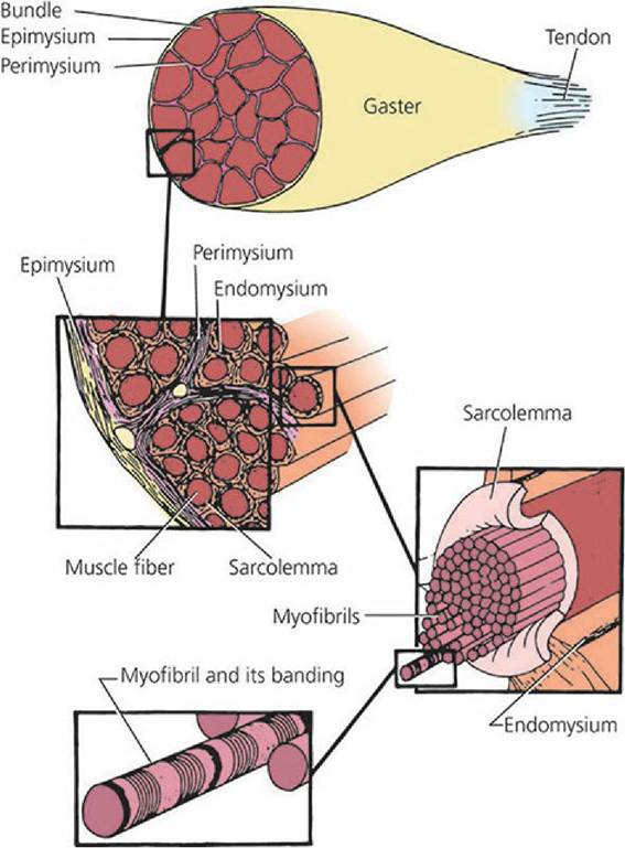
■ FIGURE 8-6 The division of muscles into smaller parts, down to myofibrils. (From Feduccia A, McCrady E. Torrey’s Morphogenesis of the Vertebrates. 5th edn. New York: John Wiley & Sons, 1991.)
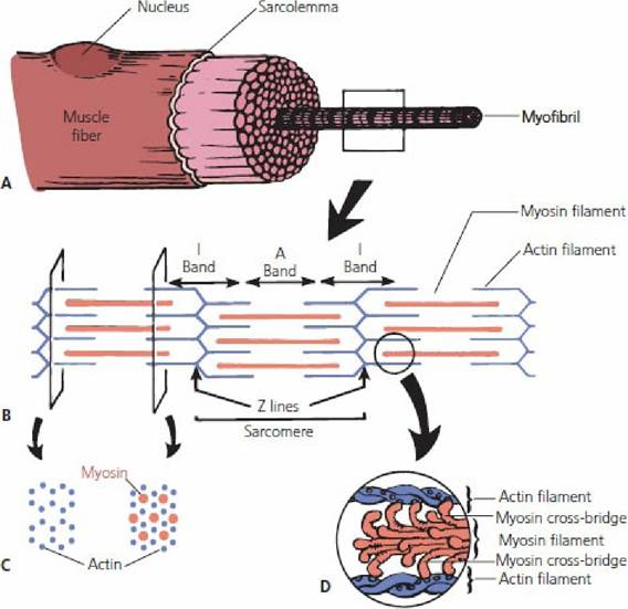
■ FIGURE 8-7 The division of myofibrils into sarcomeres. A. Cross-section of a muscle fiber. B. Longitudinal arrangement of myofilaments within sarcomeres. C. Spatial arrangement of the myofilaments within a sarcomere. D. Further details of the relationship between actin and myosin molecules.
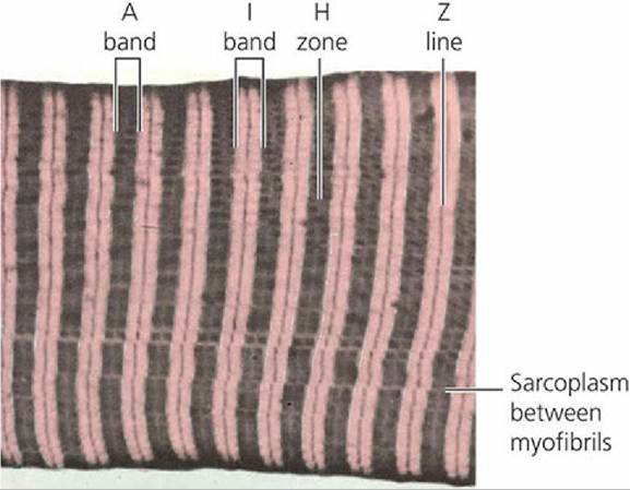
■ FIGURE 8-8 Photomicrograph of a longitudinal section of a skeletal muscle myofibril, showing the characteristic banding.
The thick dark vertical stripes are A bands of myofibrils; the light stripes contain the I bands centered by Z lines. The H zone, a paler region, is seen in the center of each A band. The pale fine lines running horizontally through the dark A bands are narrow regions of sarcoplasm lying between individual myofibrils. (From Ham AW. Histology. 7th ed. Philadelphia, PA: JB Lippincott, 1974.)Sarcotubular System
Skeletal muscle fibers contain a network of tubules known as the sarcotubular system. These tubules are located within the muscle fiber, but are outside the myofibrils. The sarcotubular system is composed of two separate tubule sets, with each set having a different arrangement among the myofibrils (Figure 8-9). The tubules that are arranged parallel to the myofibrils and encircle them are known as the sarcoplasmic reticulum. The tubules arranged transversely (right angles) to the myofibrils are known as the T tubules. T tubules extend transversely from one side of the fiber to the other. They open to the outside of the fiber (surface of the sarcolemma), and therefore their lumens contain extracellular fluid. The T tubule openings are regularly spaced throughout the length of the muscle fiber because of their orientation to each sarcomere. Similarly, their openings are regularly spaced around the circumference of the fiber so that all myofibrils are intimately served by the sarcotubular system.
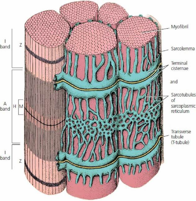
■ FIGURE 8-9 Cross-section of part of a mammalian skeletal muscle fiber, showing the sarcoplasmic reticulum that surrounds myofibrils. Two transverse (T) tubules supply a sarcomere and are in close association with the sarcoplasmic reticulum. The T tubules open to the surface of the sarcolemma and contain extracellular fluid. (From Ham AW, Cormack DC. Histology. 8th edn. Philadelphia, PA: JB Lippincott, 1979.)
In reference to a sarcomere, the T tubules are located near the junction of the actin filaments with the myosin filaments.
Therefore, each sarcomere is close to two T tubules (see Figure 8-9). The individual tubules (sarcotubules) of the sarcoplasmic reticulum are located regularly throughout the length of the muscle fiber between the T tubules, and they in turn contain intracellular fluid. The T tubules do not open into the sarcoplasmic reticulum; instead, the bulbous ends of the sarcoplasmic reticulum are closely associated with the T tubules (Figure 8-10). The point of closeness of a T tubule with the bulbous ends of two adjoining sarcoplasmic reticula is known as a triad. The principal function of the sarcotubular system is to provide a means for conduction of an impulse from the surface of the muscle fiber to its innermost aspects. The sarcoplasmic reticulum is an important storage site for calcium ions and has an important role in the initiation and termination of muscle contraction. It has an anastomosing channel-like structure that surrounds each myofibril (see Figure 8-9).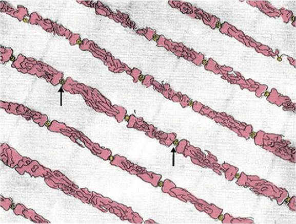
■ FIGURE 8-10 Sarcoplasmic reticulum in the extracellular spaces between the myofibrils, showing a longitudinal system paralleling the myofibrils. Also shown in cross-section are T tubules (arrows) that lead to the exterior of the fiber membrane and are important for conducting the electrical signal into the center of the muscle fiber. (From Fawcett DW: The Cell. Philadelphia, PA: WB Saunders, 1981.)
Neuromuscular Junction
Each skeletal muscle fiber is provided with one specialized area, the neuromuscular junction. This junction is the intimate association of the terminal branch of a nerve fiber with the muscle fiber (Figure 8-11). The nerve fiber ending is not continuous with the muscle fiber; there is a space between the nerve and muscle fiber. This space is centrally located on the surface of the muscle fiber. A nerve fiber can have a number of terminal branches, with each one going to a separate muscle fiber (Figure 8-12).
A motor unit consists of a nerve fiber and the muscle fibers that it innervates. A motor unit ratio of 1:150 means that one nerve fiber is innervating 150 muscle fibers, whereas a ratio of 1:4 means that one nerve fiber is innervating four muscle fibers. Hypothetically, a smaller ratio is helpful if greater precision is required for muscle contraction.
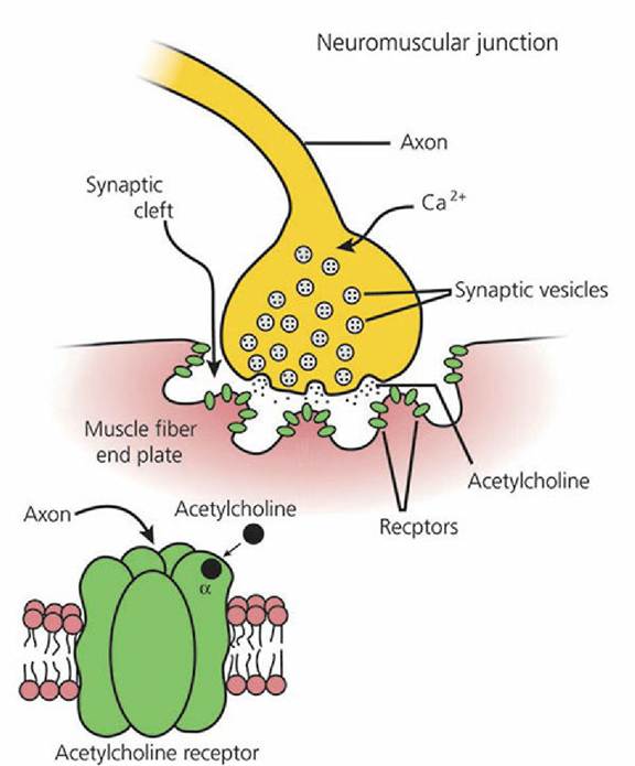
■ FIGURE 8-11 Schematic of the neuromuscular junction and the associated acetylcholine receptor channel. The terminal branch of the axon is separated from the muscle fiber by a gap known as the synaptic cleft. A neurotransmitter, acetylcholine (Ach) is stored in the membranebound synaptic vesicles. (From Bailey JG. Muscle physiology. In: Reece WO, ed. Dukes’ Physiology of Domestic Animals, 12th edn. Ithaca, NY: Cornell University Press, 2004. Used with permission of the publisher, Cornell University Press.)
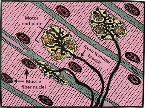
■ FIGURE 8-12 Drawing of three motor end plates synapsing on skeletal muscle fibers. Each of three terminal branches of an axon ends in a perfusion of short branches collectively designated an end plate. The bottom end plate is viewed from its edge. (From Eurell JA, Frappier BL. Dellmann’s Textbook of Veterinary Histology. 6th edn. Ames, IA: Blackwell Publishing, 2006.)
■