Motility of the Esophagus Propels Food from the Pharynx to the Stomach
The esophagus, as with other tubular portions of the gut, contains an outer longitudinal and inner circular layer of muscle. The esophagus is unique compared with other areas of the gut in that much of its muscular wall is composed of striated skeletal muscle fibers.
In most domestic animals the entire length of esophageal musculature is striated. In horses, primates, and cats, however, a portion of the distal esophagus is smooth muscle. The striated muscle portions of esophagus are under control of somatic (not parasympathetic) motor neurons in the vagus nerve, whereas the smooth muscle portions are under direct control of the ENS and indirect control of the autonomic nervous system. A myenteric plexus exists throughout the entire length of the esophagus. In the area of striated muscle, the myenteric plexus probably serves a sensory function and acts to coordinate the movements of the striated muscle portion with the esophageal smooth muscle segments and stomach.In terms of motor activity, the esophagus may be viewed as consisting of an upper sphincter, body, and lower sphincter. The upper esophageal sphincter is called the cricopharyngeal muscle. This muscle and the upper end of the esophagus are attached to the cricoid cartilage of the larynx. When deglutition is not taking place, the muscle compresses the end of the esophagus against the cartilage of the larynx, tightly closing the upper esophageal opening. During deglutition the cricopharyngeal muscle relaxes and the larynx is pulled forward. The ventral portion of the upper end of the esophagus is attached to the larynx and the dorsal portion to the cervical spine. Because of these attachments, the forward motion of the larynx in conjunction with the relatively fixed nature of the cervical spine tends to pull open the upper esophageal orifice passively (Figure 28-4).
The body of the esophagus serves as a relatively simple conduit, rapidly transferring food from the pharynx to the stomach.
Food is propelled through the esophagus by propulsive movements known as peristalsis. Peristalsis consists of a moving ring of constriction in the wall of a tubular organ. In the esophagus, these rings start at the cranial end and progress toward the stomach. The rings reduce or obliterate the esophageal lumen, thus pushing the bolus of food ahead of them in much the same manner as a person would push material out of a soft rubber tube by stripping it with the fingers. In addition to Iheconstriction of the circular muscles, there may be some contraction of longitudinal muscles just ahead of, or aboral to, the ring of circular muscle contraction. This longitudinal muscle activity increases the size of the esophageal lumen to accommodate the advancing food bolus (Figure 28-5). Peristalsis is a universal type of GI propulsive motility that exists at all levels of the gut.Duringdeglutition the upper esophageal sphincter relaxes as the pharynx constricts; food is pushed into the upper portion of the esophageal body, and a wave of peristalsis propels the material toward the stomach. As the food bolus reaches the distal end of the esophagus, the lower sphincter relaxes, and the ingested matter enters the stomach. If the esophagus
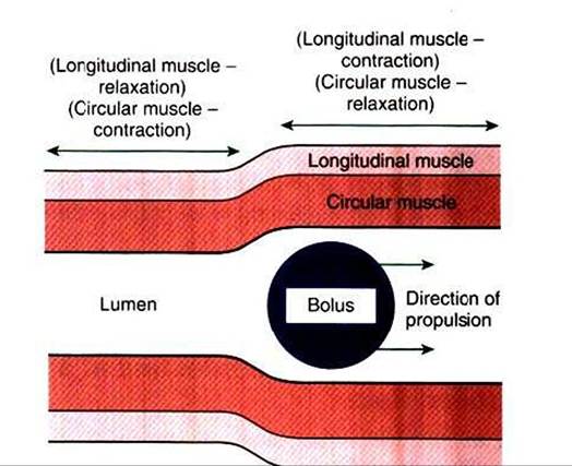
FIGURE 28-5 Peristalsis consists of a moving ring of luminal constriction preceded by an area of luminal distention.The area of constriction is created by contractions of the circular muscle, whereas the dilation is created by contractions of the longitudinal muscle. The net action is to propel a bolus of ingesta.
is not cleared of food material by the primary wave of peristalsis, secondary peristaltic waves are generated. One or more secondary waves are almost always adequate for pushing material into the stomach and clearing the esophagus. If food or foreign bodies become lodged in the esophagus, secondary waves of peristalsis may lead eventually to muscle spasms that constrict tightly around the lodged material.
These spasms frequently interfere with therapeutic attempts to remove obstructing objects in the esophagus.When deglutition is not taking place, the body of the esophagus is relaxed, but the upper and lower sphincters remain constantly constricted. The constriction of these sphincters is important because of the differences in external pressure applied to the esophagus at different points along its length. During the inspiratory phase of breathing, the portion of the esophagus within the thorax is subjected to less-than- atmospheric pressure. If the two esophageal sphincters were not tightly closed, inspiration would cause aspiration of air from the pharynx and reflux of ingesta from the stomach into the body of the esophagus, in the same manner as inspiration draws air into the lung. Stomach contents would be drawn into the esophagus because inspiratory pressures in the thorax are lower than intraabdominal pressure. It is particularly important that the lower esophageal sphincter remain closed during inspiration because the mucosa of the esophagus is not equipped to resist the caustic actions of gastric contents; thus movement of stomach contents into the esophagus would cause damage to the esophageal mucosa.
In many species the action of the lower esophageal sphincter is aided by the anatomical nature of the attachment of the esophagus and stomach. The esophagus enters the stomach obliquely, allowing distention of the stomach to block the esophageal opening in a valvelike manner. Duringdeglutition the longitudinal muscle of the esophagus contracts, shortening the esophagus and opening the valve at the junction with the stomach. This anatomical arrangement, along with the lower esophageal sphincter, is particularly well developed in the horse, making reflux of stomach material into the esophagus extremely rare in this species. In many cases, when the intragastric pressure of the horse is pathologically raised, the stomach ruptures before vomiting or esophageal reflux takes place.