Organization of the Digestive System
The digestive system (digestive tract) consists of a muscular tube lined with mucous membrane that is continuous with the external skin at the mouth and at the anus. Its primary functions are prehension, mastication, digestion, and absorption of food, and elimination of solid wastes.
The digestive system reduces the nutritious constituents of the food to molecular compounds that are small enough to be absorbed and used for energy and for building other compounds for incorporation into body tissues.Elements of the digestive system are the mouth, pharynx, esophagus, forestomach (ruminants), glandular stomach, small intestine, large intestine, rectum, and the accessory glands (salivary glands, liver, and pancreas).
Caudal to the diaphragm, the components of the digestive tract lie within the abdominal and pelvic cavities. Here they are invested with a simple squamous epithelium that is also called a mesothelium or serosa. Within these body cavities, the serosa is identified as peritoneum. Like the pleura within the thoracic cavity, it is named according to the structures to which it is applied: where it lies directly on the organ, it is called visceral peritoneum, and where it invests the abdominal wall, it is parietal peritoneum (see Fig. 1-9).
Parietal and visceral peritonea are continuous with one another through reflections of the serosa that attach the organs to the body wall.
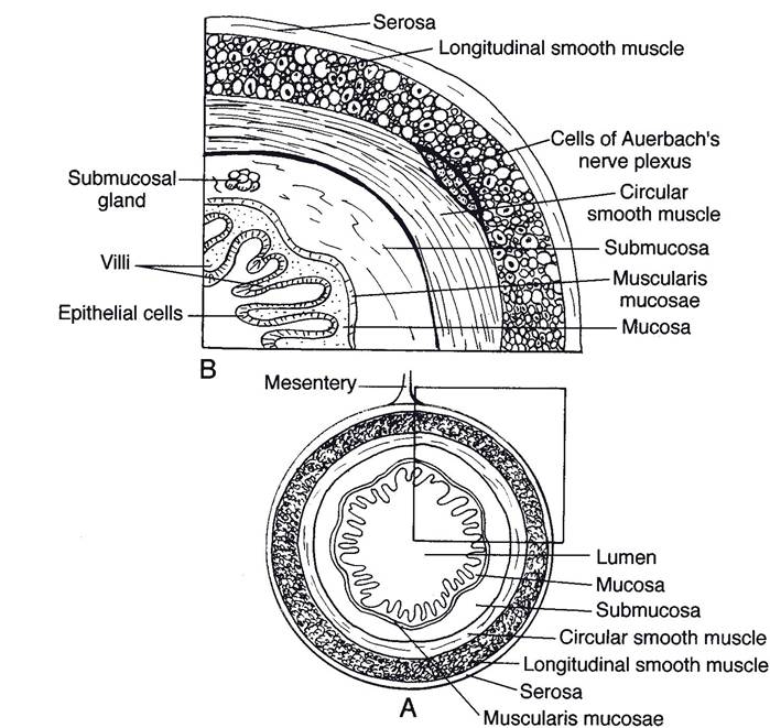
Figure 20-1. Layers of a typical segment of gut. (Reprinted with permission of Wiley-Blackwell from Reece W.O.
Physiology of Domestic Animals. 2nd ed. Baltimore: Williams & Wilkins, 1997.)
These attachments collectively constitute the mesenteries, and they are named for the organ they suspend (discussed later). Blood vessels, lymphatics, and nerves travel within the mesenteries, reaching the organs through them.
The wall of the digestive tract comprises four layers, or tunics. These are, from within outward: (1) the tunica mucosa, (2) the tunica submucosa, (3) the tunica muscularis, and (4) the tunica serosa, or (where organs lie outside body cavities) tunica adventitia (Fig. 20-1).
The tunica mucosa is the layer closest to the space (the lumen) inside the digestive tract. It has three histologic layers. The innermost layer consists of a stratified squamous epithelium from the mouth to the level of the glandular part of the stomach; from this point to the anus, the epithelium is of the simple columnar type. Underlying the epithelium of the tunica mucosa are a layer of connective tissue and a variably present smooth muscle layer.
The tunica submucosa is a layer of loose connective tissue in which are found blood vessels and nerves. in some locations, glands of the digestive tract can be found in the submucosa, as can lymphatic nodules.
As motility is important to the function of the digestive system, the tunica muscularis is generally well developed. in the horse, the cranial two-thirds of the tunica muscularis of the esophagus is striated muscle; in the pig, all but the most distal end of the esophagus is striated; and in ruminants, the entire esophagus has striated muscle. From this point and distal, the muscle cells are smooth (involuntary) and are generally arranged in two layers. The deeper layer has fibers that encircle the gut, and the more superficial muscle layer assumes a longitudinal arrangement.
The outermost tunic consists of the visceral peritoneum and scant connective tissue underlying it. This is the tunica serosa. The thoracic and cervical parts of the esophagus are not suspended directly within a body cavity (the thoracic esophagus is surrounded by the tissues of the mediastinum) and therefore do not have a tunica serosa. Instead, the surrounding connective tissue in these locations constitutes a tunica adventitia. Likewise, the distal end of the rectum and anal canal are outside the pelvic cavity and surrounded by a tunica adventitia.