Much of the general description of the teeth was based on the dentition of the dog, in which the most remarkable features are the prominence of the canine teeth and the marked regional specialization of the others (see Fig. 3.16).
The upper dental arch, despite having fewer teeth, is slightly longer than the lower one; the upper teeth therefore bite on the buccal side of the lower ones in a shearing action.
This feature precludes lateral movement of the lower jaw, making grinding impossible. There is little occlusal contact between upper and lower teeth except caudally, where some crushing of food is possible. The first few premolars do not touch at all, which creates the so-called carrying space. Dogs and cats bolt rather than chew their food.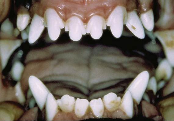
FIG. 11.21 Trilobular form of upper incisors.
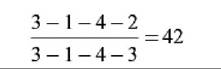
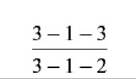
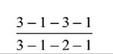
The formula for temporary dentition in dogs is
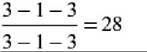
and for the permanent set is
The Triadan system is also available for reference to specific teeth. In this system, each tooth is assigned a three-digit number. The first digit (in the hundreds place) indicates the quadrant of the mouth: 1(00) indicates the right upper, 2(00) the left upper, 3(00) the left lower, and 4(00) the right lower quadrant. The other two digits indicate the place of the tooth in the dental arcade, 01 being the most mesial. Thus 102 specifies the upper right second incisor, 409 the lower right first molar.
The incisor teeth are rather loosely embedded in the incisive bones and mandible. On eruption the upper incisor crowns present a central cusp flanked by two smaller ones; the mesial cusp is
lacking on the lower incisors (Fig. 11.21). These features are lost as wear reduces the incisors to simple prismatic pegs. The wear gives some indication of a dog's age but is not very reliable because of differences in skull size, frequency of malocclusion, and individual variation in the diet and habits (Fig.
11.22). All incisors have a single root. They are mainly for nibbling, both in grooming and when detaching small morsels.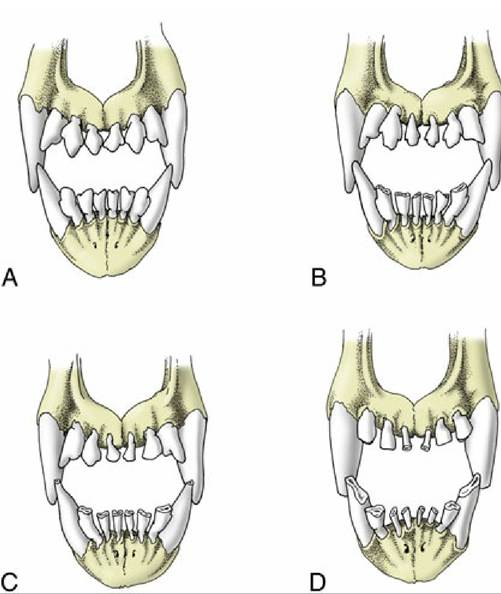
FIG. 11.22 Changes in the canine incisors with increasing age. (A) Six months; (B) about 2½ years; (C) about 6 years; (D) about 10 years.
The root of the canine is especially massive—larger indeed than the crown—and curves caudally to lie dorsal (or ventral) to the first premolar (Fig. 11.23). These teeth are occasionally removed in aggressive dogs. Simple extraction is made impossible by the size and firm implantation of the root; the attempt to draw one free risks fracture of the jaw. It is necessary to resect the bone over the lateral surface of the root before it can be elevated from its socket. Abscesses of the upper canine teeth may fistulate into the nasal cavity.
In adult dogs, there are four premolar teeth, the first of which may have either one or two roots, whereas the others have two. The one exception is the upper fourth premolar or sectorial tooth, which has three roots (Fig. 11.24). The four premolars increase in size and complexity from the first to last in both jaws. The laterally compressed crowns are triangular in profile, presenting small mesial and distal cusps to each side of the principal one. The last upper premolar, P4, is massive and has a small medial part, with its own root, which encroaches on the hard palate. The molars decrease in size from first to last. The two upper molars, though still tuberculate, have flatter crowns than the premolars and are orientated transversely rather than rostrocaudally (see Fig. 11.24). They have three diverging roots. The first of the lower molars, M1, the sectorial tooth, is the largest in the lower series. It is flattened from side to side and has two thick divergent roots that occupy most of the width of the jaw. Extraction must be performed carefully to avoid fracture of the mandible.
M2 and M3 are much smaller; they engage the last upper molar and, like it, have flat tuberculate crowns. They also have two roots each.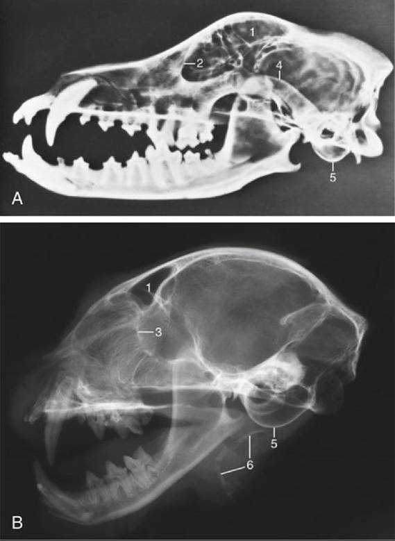
FIG. 11.23 (A) Radiograph of half of a canine skull showing the permanent teeth and their roots. (B) Radiograph of half of a feline head. 1, Frontal sinus; 2, orbital rim; 3, cribriform plate; 4, zygomatic arch; 5, tympanic bulla; 6, hyoid apparatus.
It is important to know the pattern of the sockets to ensure that no part is left behind after extraction of a tooth (see Fig. 11.24). Multiple roots always diverge, and it is frequently necessary to split a tooth before it can be extracted to avoid causing excessive trauma.
Brachygnathic breeds often have less than the full complement of teeth: upper and lower P1 and M3 are those most often missing. The cheek teeth of these breeds may be more obliquely placed than normal to fit in the foreshortened jaws.
At birth, a puppy is toothless. The first teeth appear within a few weeks, and the deciduous set is complete and functional by the end of the second month. The first replacement tooth erupts after a further month, or little more, and the permanent set is complete by the sixth or seventh month, a remarkably early age (Table 11.1). Permanent teeth erupt earlier in larger breeds of dogs. The temporary teeth in general resemble those of the definitive set but are smaller and sharper. They have long slender roots. A temporary canine is sometimes retained after the replacement tooth has erupted because the latter appears beside its predecessor and produces asymmetrical and sometimes insufficient resorption pressure. In such cases the temporary canine is found caudal to its replacement in the upper jaw and lateral to it in the lower jaw. Retained teeth should be removed to allow their replacements to attain their normal positions. The three temporary premolars are properly designated p2, p3, and p4; the tooth known as the first premolar erupts several weeks later than these and is part of the permanent dentition (Table 11.1).
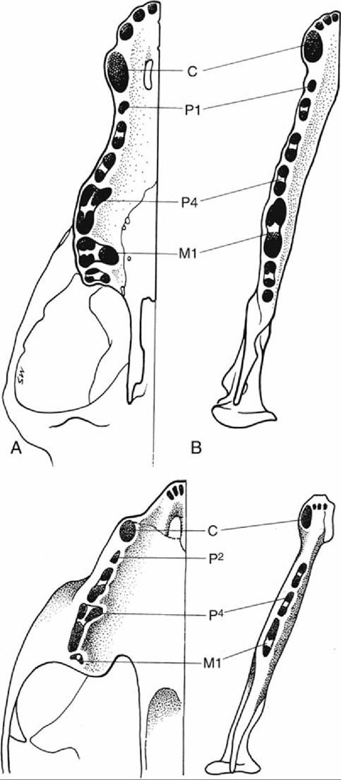
FIG. 11.24 The tooth sockets in (top) canine and (bottom) feline (A) upper and (B) lower jaws to show the number and disposition of the roots. C, canine; M, molar; P, premolar; numbers indicate tooth positions.
The upper teeth are innervated by the infraorbital nerve, and the rostral members of the series can be desensitized by blocking of the nerve within the infraorbital foramen. The lower teeth are supplied by the inferior alveolar nerve, which can be blocked at a site a centimeter or so caudal to the last tooth, before it enters the mandible. The rostral members of this series can also be desensitized by blocking of the nerve within the mental foramen.
» TABLE 11.1
Eruption Dates of the Dog's Teeth
* Permanent teeth erupt slightly earlier in large breeds.
Modified from Schummer A, Nickel R, Sack WO: The viscera of the domestic mammals, ed 2, New York, 1979, Springer-Verlag; and Evans HE: Miller’s anatomy of the dog, ed 3, Philadelphia, 1993, Saunders.
The cat has sharp and pointed teeth. The formula for the temporary dentition reads
and for the permanent dentition reads
The smaller number of cheek teeth is due to the absence of P1 and M2 and of P1, P2, M2, and M3 (see Fig. 3.17). The molar loss deprives the cat of flat-crowned crushing teeth, leaving an exclusively shearing bite (Fig. 11.25). P4, the upper sectorial, is the only tooth to have three roots, which are implanted only a few millimeters from the ventral wall of the orbit. Its lower counterpart is M1. It is not uncommon to find that one or more of the smaller incisor teeth have been shed by the time cats settle into middle age, without obvious cause.
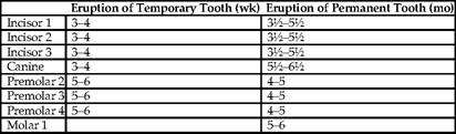
In kittens, eruption of deciduous teeth typically begins during the third postnatal week. The permanent teeth are all in place by about 6 months of age. However, there is so much individual and breed variation that the average eruption and replacement dates given in Table 11.2 are unreliable guides to age.
Plaque deposition and consequent periodontal disease are common in both companion species. In cats, such disease is often accompanied by resorptive lesions at the necks of the teeth.
» TABLE 11.2
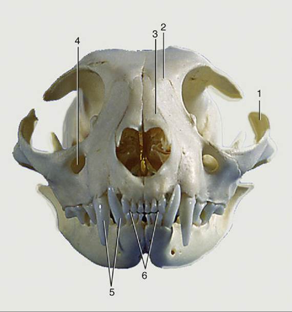
FIG. 11.25 Feline skull, rostral view. 1, Zygomatic arch; 2, frontal bone; 3, nasal bones; 4, infraorbital foramen; 5, upper and lower canine teeth; 6, upper and lower incisors. Upper teeth are in incisive bones and lower teeth in the mandible.
Eruption Dates of the Cat's Teeth
From Schummer A, Nickel R, Sack WO: The viscera of the domestic mammals, ed 2, New York, 1979, Springer-Verlag.