The articular surfaces of the temporomandibular joint are nearly congruent.
The transverse cylinder provided by the mandible fits within a trough on the undersurface of the zygomatic process of the temporal bone (Figs. 11.23, 11.26, 11.28, and 11.29). The trough is enlarged caudally by a prominent retroarticular process that securely cups the cylinder and prevents its luxation in a caudal direction.
In keeping with the congruence of the joint, the articular disk is thin. The joint capsule is strengthened by a lateral ligament.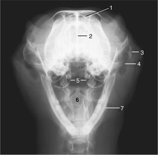
FIG. 11.26 Rostrocaudal open-mouth radiograph of a feline head. 1, Frontal sinus; 2, nasal septum; 3, zygomatic arch; 4, temporomandibular joint; 5, tympanic bullae; 6, axis with dens; 7, mandible.
Movement of the mandible is almost exclusively of a hinge nature, but slight protrusion is possible when the mouth is fully open. Lateral movement may be produced by trauma and occasionally is so severe that the coronoid process engages the zygomatic arch, locking the jaws in the depressed position.
The joint lies under cover of the caudal part of the masseter, where the dorsal buccal branch of the facial nerve crosses the border of the muscle. It is rostral to the parotid gland.
The masticatory muscles have been sufficiently described (p. 105-106).