Muscle Stretch and Action Potentials Along Spindle Sensory Neurons Lead to Reflex Contraction of the Extrafusal Muscle Fibers
The sensory output neurons of the muscle spindle enter the CNS, where they make excitatory, monosynaptic connections with α motor neurons that return to the extrafusal fibers of the same muscle (see Figure 8-3).
Therefore, stretching a given muscle can lead to a rapid, reflex contraction of that same muscle, bringing it back to its original length. Stretching the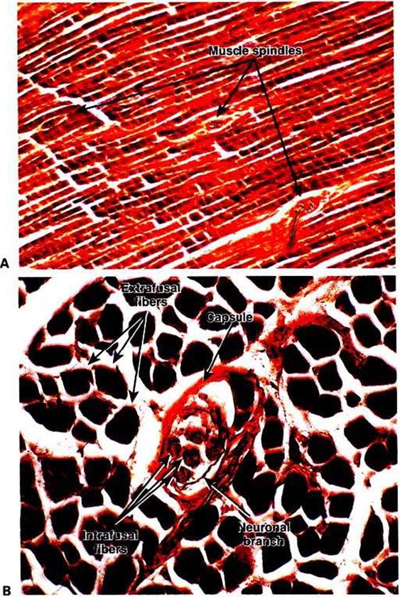
FIGURE 8-2 The muscle spindle receptor is an encapsulated group of specialized (intrafusal) skeletal muscle fibers supplied with both motor and sensory innervation. Ar Longitudinal section through a skeletal muscle showing that the encapsulated muscle spindles are oriented parallel to the more numerous e?trafusal fibers of the muscle.The ends of the muscle spindle are attached to the extracellular matrix of the e?trafusal fibers. B, Higher- magnification view of a transverse section through a muscle spindle. Intrafusal fibers can be seen within the spindle's tissue capsule.These fibers are fewer, shorter, and more slender than the surrounding e?trafusal fibers. A portion of the spindle's innervation can also be seen. (Images courtesy Dr.Tom Cacecir Department of Biomedical Sciences and Pathobiologyr College OfVeterinary Medicine, VirginiaTech.)
muscle lengthens the intrafusal muscle fibers of the spindle, increasing the frequency of action potential discharge along the sensory output neurons of the spindle. This leads to an increase in action potential frequency in the α motor neurons on which the spindle sensory neurons synapse. This produces contraction of the extrafusal fibers innervated by those α motor neurons, which results in contraction (shortening) of the muscle. Contraction of the muscle results in a shortening of the muscle spindle’s equatorial region.
This eventually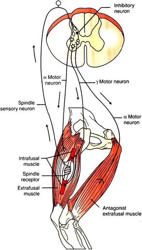
FIGURE 8∙3 The muscle spindle stretch reflex (illustrated here as the "knee jerk reflex") begins when the spindle receptor organ is Stretched-This causes action potentials on the receptor's sensory neurons, which in turn cause excitatory postsynaptic potentials on the α motor neurons returning to the extrafusal muscle fibers of that same muscle. Action potentials on the α motor neurons cause extrafusal muscle fibers to contract, and the knee extends ("jerks").Through an inhibitory interneuron, the «motor neurons to the antagonist muscles are simultaneously inhibited.
reduces the frequency of action potentials occurring on the spindle sensory neurons to the prestretch level, terminating the response. (The cycle is a classic negative-feedback system.)
The reflex just described can be elicited by striking the patellar tendon (insertion tendon of quadriceps muscle) with a blunt object. Because this tendon goes over a “pulley” (the patella), hitting this tendon results in a longitudinal stretch of the whole quadriceps muscle, thus also stretching the muscle spindles. Action potentials from spindle sensory neurons go to the lumbar spinal cord, by way of the dorsal roots, and cause excitatory postsynaptic potentials (EPSPs) on the α motor neurons of the motor units that return to the quadriceps muscle.
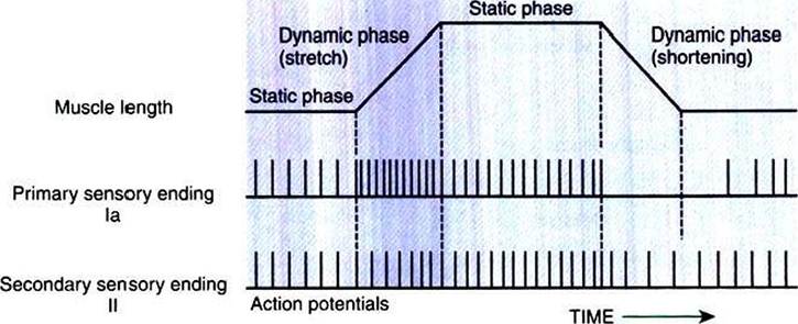
FIGURE 8-4 The muscle spindle can signal the steady-state length of the muscle, as well as the onset and velocity of stretch. When the muscle is stretched, both type Ia and type Il spindle sensory neurons have a higher action-potential firing rate at the muscle's new static length. During the dynamic stretching phase, the action-potential firing rate of the type Ia spindle sensory neuron increases rapidly and in proportion to the velocity of stretch.
Spindle sensory neurons can also register a decreased steady-state length of the muscle, but type Ia and type Il neurons display differential sensitivity to the dynamic phase of shortening (From Brodal P: The central nervous system: structure and function, ed 2, NewYork, 1998, Oxford University Press.)This causes contraction of the quadriceps muscle and an extension of the knee joint and is an example of the muscle stretch reflex, or myotatic reflex. When it is applied to the quadriceps muscle, it is called the knee jerk reflex, but the mechanisms are present in almost all muscles. However, this is the muscle from which it is easiest to evoke the stretch reflex, because it is one of the few whose tendon goes over a sesamoid pulley before inserting on the next bone. Because of the pulley under the tendon, a lateral deflection of the tendon, as from a reflex hammer, results in a longitudinal stretch of the muscle and thus the reflex. Hitting other tendons only moves the muscle belly laterally and does not easily result in the stretch reflex. Therefore, in the clinical neurological examination of most animals, the kneejerk reflex is the most commonly evoked muscle stretch reflex.
The muscle spindle organ and stretch reflex allow the CNS to make automatic, usually unconscious adjustments to muscle stretch imposed by small changes in body position or the weighting of a muscle. Such adjustments can return the muscle to its original length, often returning a joint to its original position. Clinical examination of the stretch reflex provides clues about the peripheral or CNS integrity of its sensory and motor components.
When the stretch reflex acts to return a joint to its original position, the antagonist of the stretched muscle must relax in order for the joint to be able to move. Therefore, in the stretch reflex, some terminal branches of individual spindle sensory neurons do not synapse directly on the α motor neurons of the stretched muscle, but rather synapse on inhibitory spinal interneurons (see Figure 8-3). These neurons, which lie completely within the spinal cord, are also activated by the stretch of the muscle. When they fire action potentials, however, they cause inhibitory postsynaptic potentials (IPSPs) on α motor neurons that innervate the antagonist of the stretched muscle. This prevents the contraction of the antagonistic muscle.