The Central Nervous System Can Control Spindle Sensitivity Directly Through the Gamma (γ) Motor Neurons
Contraction of the extrafusal muscle fibers is controlled by the larger α motor neurons; intrafusal muscle fibers of the spindle are controlled by the smaller γ motor neurons.
The γ motor neurons innervate the intrafusal muscle fibers at their polar ends (see Figures 8-1 and 8-3), the regions containing contractile protein. Action potentials on the γ motor neurons cause shortening of the polar regions of the intrafusal muscle fibers, stretching the equatorial portion.An important function for this motor innervation of a receptor organ is to regulate the sensitivity of the muscle spindle. Shortening of a gross muscle resulting from initiation of extrafusal muscle fiber contraction has the potential to slacken the intrafusal muscle fibers given their parallel relationship to the extrafusal fibers. I his would severely limit the ability of the muscle spindle to transduce stretch. However, this does not normally occur because contraction of the polar regions of intrafusal fibers resulting from γ motor neuron activation is initiated concurrently with shortening of extrafusal fibers caused by α motor neuron activation. I his allows the spindle receptor organ to remain taut and sensitive to sudden stretches of the gross muscle over the entire range of its length. This γ motor neuron control mechanism can also function to differentially regulate the sensitivity of the muscle spindle, depending on the type of movement to be made (e.g., novel and unpredictable vs. stereotypical). Chapter 10 describes how co-activation of both the α and γ motor neurons allows the brain to test whether the amount of contraction intended by the brain was what actually occurred.
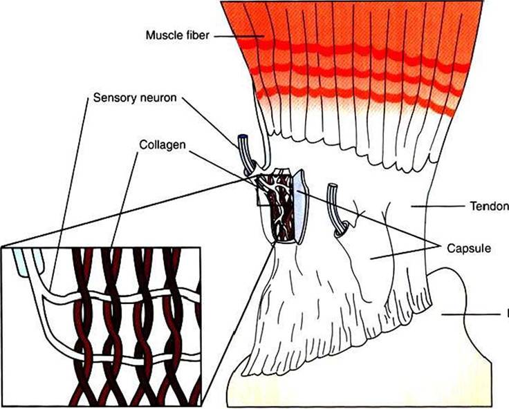
FIGURE 8-5 The Golgi tendon organ is located in the tendons of skeletal muscle, in series with the extrafusal fibers. It detects tension in the tendon, produced by muscle contraction, and sends information about this tension to the central nervous system. Sensory neuron branches of the organ are intertwined among braided collagen fibrils (inset), which fold up and pinch the neural branches when tension develops in the tendon.