Muscles of the Pelvic Limb
The hip is a ball-and-socket joint and as such can move in nearly any direction (Fig. 7-10). However, the chief movements are extension and flexion of the femur. Adduction and abduction are also fairly common movements, and some rotation is possible.
See Table 7-2 for a list of muscles of the pelvic limb.
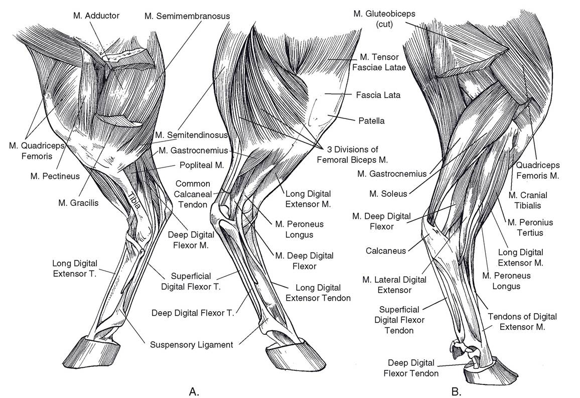
Figure 7-10. Muscles of the pelvic limb. A) Equine. Left) medial view. Right) lateral view. B) Bovine: lateral view.
Table 7-2. Muscles of the Pelvic Limb
| Muscle | Origin | Insertion | Primary Actions | Innervation | |
| m. biceps femoris (m. gluteobiceps in ruminants) | Sacrum, sacrosciatic ligament, ischial tuber | Patellar ligament, patella, & tibia via femoral & crural fascia, calcaneal tuber | Extension of hip & stifle (cranial part of m.); flexion of stifle & extension of hock (caudal part) | Caudal gluteal & tibial nn. | |
| m. semitendinosus | Sacrum & caudal vertebrae, sacrosciatic ligament, ischial tuber | Cranial tibia, crural fascia, calcaneal tuber | Extension of hip, stifle, & hock (when weight-bearing); flexion of stifle (when limb off ground) | Caudal gluteal & tibial nn. | |
| m. semimembranosus | Caudal vertebrae, sacrosciatic ligament, ischial tuber | Medial distal femur, medial proximal tibia | Extension of hip & stifle (when weightbearing); flexion of stifle (when limb off ground) | Caudal gluteal & tibial nn. | |
| m. iliopsoas | Ventral aspects of lumbar vertebrae, ilium, sacrum | Lesser trochanter of femur | Flexion of hip | Ventral branches of lumbar spinal nn. | |
| m. sartorius | Ilium | Medial fascia of stifle | Flexion of hip, extension of stifle | Femoral n. | |
| m. quadriceps femoris m. rectus femoris m. vastus lateralis m. vastus intermedius m. vastus medialis | Proximal femur (vastus mm), body of ilium (m. rectus femoris) | Patella | Extension of stifle, flexion of hip (rectus only) | Femoral n. | |
| m. tensor fasciae latae | Tuber coxae | Fascia lata | Flexion of hip, extension of stifle | Cranial gluteal m. | |
| m. gluteus superficialis | Gluteal fascia | Third trochanter of femur | Abduction of hip | Cranial & caudal gluteal nn. | |
| m. gluteus medius | Wing of ileum, tuber coxae, sacrum, sacrosciatic ligament | Greater trochanter of femur | Extension of hip | Cranial gluteal n. | |
| m. gluteus profundus | Body of ilium | Greater trochanter of femur | Abduction of hip | Cranial gluteal n. | |
| m. gracilis | Pelvic symphysis | Medial aspect tibial crest | Adduction of limb | Obturator n. | |
| m. pectineus | Pubis | Medial aspect proximal tibia | Adduction of limb, flexion of hip | Obturator n. | |
| m. adductor | Ventral pelvis | Medial femur | Adduction of limb | Obturator n. | |
| Table 7-2. Continued | |||||
| Muscle | Origin | Insertion | Primary Actions | Innervation | |
| m. quadratus femoris | Ventral sacrum | Caudal proximal femur | Outward rotation of hip, hip extension | Sciatic n. | |
| External obturator m. (m. obturatorius externus) | Ventral pelvis, covering obturator foramen | Trochanteric fossa of proximal femur | Outward rotation of hip, limb adduction | Obturator n. | |
| Internal obturator m. (m. obturatorius internus) | (Absent in ruminants) covering obturator foramen inside pelvic canal | Trochanteric fossa of proximal femur | Outward rotation of hip, hip extension | Sciatic n. | |
| mm. gemelli | Ischium | Trochanteric fossa of proximal femur | Outward rotation of hip, hip extension | Sciatic n. | |
| m. popliteus | Lateral aspect of lateral femoral condyle | Caudoproximal tibia | Stifle flexion | Tibial n. | |
| m. gastrocnemius | Caudal aspect of femoral condyles | Tuber calcanei | Hock extension | Tibial n. | |
| m. flexor digitorum superficialis (superficial digital flexor m.) | Caudodistal femur, between heads of m. Gastrocnemius | Tuber calcanei, plantar aspect of middle phalanx | Hock extension, digital flexion | Tibial n. | |
| m. tibialis cranialis | Craniolateral aspect proximal tibia | Medial tarsus & metatarsus | Flexion of hock | Peroneal n. | |
| m. peroneus tertius | Extensor fossa of distolateral femur | Cranial aspect of distal tarsus & proximal metatarsus | Flexion of hock | Peroneal n. | |
| m. peroneus longus | Head of fibula & lateral femoral condyle | Plantar aspect of distal tarsus & metatarsus | Flexion of hock | Peroneal n. | |
| m. extensor digitorum longus (long digital extensor m.) | Extensor fossa of distolateral femur | Dorsal aspect of distal phalanx | Flexion of hock, extension of digits | Peroneal n. | |
| m. extensor digitorum lateralis (lateral digital extensor m.) | Proximal fibula | Middle phalanx (artiodactyls), tendon of long digital extensor (Equidae) | Flexion of hock, extension of digits | Peroneal n. | |
| m. flexor digitorum profundus (deep digital flexor m.) | Proximocaudal tibia | Plantar aspect distal phalanx | Extension of hock, flexion of digits | Tibial n. | |
Muscles Acting on the Hip Joint
Extensors of the Hip. The chief extensors of the hip are the so-called hamstring muscles, which pass caudal to the hip from the tuber ischiadicum to the proximal end of the tibia, fibula, or calcaneus of the tarsus. They include the m. biceps femoris (the most lateral of the caudal muscles of the thigh), the m. semitendinosus (the middle muscle of the caudal group), and the m. semimembranosus (the medial muscle of this group). The divisions between these muscles can be seen as vertical grooves in animals that are not very fat. in the horse, the m. bicepsfemoris and m. semitendinosus extend dorsad over the rump to attach to the sacral and caudal (coccygeal) vertebral spines (the so- called vertebral heads of these muscles). in most other animals, the hamstring muscles originate almost exclusively from the tuber ischiadicum. In ruminants, the m. biceps femoris is blended with the superficial gluteal muscle and is therefore called the m. gluteobiceps. This combined muscle is a powerful extensor of the hip, stifle, and hock.
The middle gluteal muscle (m. gluteus medius) is another strong extensor of the hip. it originates from the wing of the ilium and inserts on the greater trochanter of the femur, a lever projecting above the hip.
Flexors of the Hip. Flexors of the hip are cranial to the femur. The most important are the m. iliacus and m. psoas major, which insert on the lesser trochanter on the medial side of the femur. Together they are called the iliopsoas muscle. The m. iliacus originates from the ventral surface of the wing of the ilium. The m. psoas major originates from the ventral surfaces of the lumbar transverse processes.
The m. sartorius is a thin, straplike muscle that extends from the tuber coxae to the tibia, diagonally crossing the medial surface of the thigh. The m. rectus femoris (one head of the m. quadriceps femoris) and the m. tensor fasciae latae also flex the hip and are also described as extensors of the stifle.
Abductors of the Hip. Abductors of the hip extend laterally over the hip joint so as to move the limb away from the median plane. The deep gluteal muscle (m. gluteus profundus) extends from the spine of the ischium laterad over the hip joint to insert on the greater trochanter.
The superficial gluteal muscle (m. gluteus superficialis) extends from the sacral vertebral spines to the third trochanter just distal to the greater trochanter. The m. tensor fasciae latae extends from the tuber coxae to the lateral femoral fascia, which attaches to the patella. in addition to abducting the hip joint, this muscle flexes the hip joint and extends the stifle.
Adductors and Rotators of the Hip. Adductors of the hip pull the limb toward the median plane. They are all on the medial aspect of the thigh, extending from the pelvis to either the femur or the tibia. The m. gracilis is the most medial muscle extending from the symphysis of the pelvis to the tibia.
The m. pectineus, a small spindle-shaped muscle deep to the m. gracilis, is both an adductor and flexor of the hip.
The m. adductor is the largest muscle on the medial side of the thigh. it extends from the ventral aspect of the pelvis to the medial side of the femur and tibia. it is a strong adductor but may also help to extend the hip.
The m. quadratus femoris is an adductor of the thigh. several other small muscles in this deep layer of hip musculature extending from the area of the obturator foramen are outward rotators of the thigh. They include the internal and external obturator muscles (mm. obturatorius internus et externus), and the mm. gemelli. Ruminants lack the internal obturator muscle.
Muscles Acting on the Stifle
The stifle is essentially a hinge joint, so the muscles acting on it are either extensors or flexors.
Extensors of the Stifle. One large muscle, the m. quadriceps femoris, is the primary extensor of the stifle. This muscle has four heads; their distinct origins and clearly distinguishable muscle bellies make it common practice to name them as separate muscles. The longest head, the m. rectus femoris, originates from the ilium just above the acetabulum. The other three heads, m. vastus medialis, m. vastus intermedius, and m. vastus lateralis, originate from the respective areas of the shaft of the femur. All four heads insert on the patella. The patella, being fastened to the front of the tibia by the patellar ligaments, extends the stifle when it is pulled proximad by the m. quadriceps femoris. Because of its origin on the ilium, the m. rectus femoris also flexes the hip.
Flexors of the Stifle. The chief flexors of the stifle are the hamstring muscles, which also extend the hip (m. biceps femoris, m. semitendinosus, m. semimembranosus; discussed earlier). In addition, the extensor muscles of the hock that originate on the caudal surface of the distal end of the femur may also flex the stifle. These muscles include the m. gastrocnemius and the superficial digital flexor m. (discussed later). The m. popliteus is a relatively small muscle caudal to the stifle. Its chief action is flexion of the stifle, although it may slightly rotate the leg (tibia and fibula) mediad.
Muscles Acting on the Hock
The principal actions of the hock are extension and flexion.
Extensors of the Hock. Extensors of the hock primarily attach to the calcaneus (point of the hock) by way of the common calcanean tendon. The m. gastrocnemius and superficial digital flexor m. originate from the caudal aspect of the distal femur, and their tendons make up the bulk of the common calcanean tendon. They are joined in part by portions of the m. biceps femoris, m. gracilis, and m. semitendinosus, which also assist in extending the hock and hip and flexing the stifle. The deep digital flexor m. also extends the hock.
Flexors of the Hock. Flexors of the hock include the m. tibialis cranialis and the various peroneal muscles, whose tendons pass over the dorsal surface of the hock to insert on the tarsus and metatarsus. The m. peroneus tertius is the only named peroneal muscle in the horse. Additionally, the m. peroneus longus is found on the ox, sheep, goat, pig, and dog. The m. peroneus brevis is found only in carnivores and humans. The digital extensors also flex the hock, because their tendons pass over its flexor surface.
Muscles Acting on the Digit
Digital extensors of the thoracic limb also tend to extend the carpus, while digital extensors of the pelvic limb tend to flex the hock. Likewise, digital flexors of the thoracic limb produce flexion of the carpus, while digital flexors of the pelvic limb produce extension the hock. Muscles acting on the digits of the pelvic limb are similar in arrangement to those of the thoracic limb.
Extensors of the Digit. The long digital extensor muscle (m. extensor digitorum longus) originates from the distal end of the femur and passes distad to insert on the distal phalanx of each digit. As with the common extensor in the thoracic limb, the tendon has one part in the horse; two parts in the ox, goat, and sheep; and four parts in the pig and carnivores.
The lateral digital extensor muscle (m. extensor digitorum lateralis) lies between the extensor and flexor groups of muscles of the crus. In the horse, its tendon joins that of the long digital extensor m. at about the middle of the cannon. In ruminants, it inserts via a separate tendon on the proximal middle phalanx of the fourth digit. In pigs, it resembles the ruminant condition, with the addition of a small deep part of the muscle that features a separate tendon inserting on the fifth digit.
Flexors of the Digit. The superficial and deep digital flexor mm. are arranged in the pelvic limb similarly to the thoracic limb. However, the tendon of the superficial digital flexor muscle also attaches to the calcaneus.