Muscular System
The second of two complementary systems comprising what we recognize as the Iocomotory complex is the muscular system. We will be concentrating on what most of us recognize as “muscle, ” in ourselves and other vertebrates, i.e., the bulk of our body mass that gives our bodies shape and definition.
The function of muscle is contraction. In vertebrates, there are three types of muscle, smooth or involuntary, associated with systems not involved in body locomotion, e.g., the digestive system. Cardiac muscle, involuntary in function, occurs only in the heart. Skeletal or striated muscle which is voluntary in function is generally associated with visible body movements. It also aids in such activities as returning venous blood and lymphatic fluid, especially from the posterior limbs, toward the heart.
The tissues of the muscular system bring about not only externally visible locomotory movements, but also some important subtle, sophisticated, internal movements, such as food propulsion through the digestive tract, adjustment of blood vessel diameters to control blood volumes in various body regions, regulation of respiratory tube diameter, erection of hair follicles, and intrinsic eye functions such as dilation and constriction of the pupil.
The gross anatomy of a skeletal muscle includes the swollen middle region called the belly, the less movable end known as the origin, and the more movable end called the insertion [Figure 2-1]. In some muscles that are capable of several actions, the origin and insertion may be reversed during contraction. The points of origin and insertion are marked by the presence of dense connective tissue which anchor each muscle to a bone, straplike tendons, or to other muscles, by means of flat sheetlike aponeuroses.
Macroscopically, the structure of a muscle consists of a variable number of muscle cells (fibers) each encased in connective tissue, the endomysium, occurring in bundles called fasciculi and wrapped in connective tissue, the perimysium.
Finally, groups of fasciculi surrounded by connective tissue, the epimysium, make up the whole muscle, e.g., the Biceps brachii. Microscopically, each of the muscle fibers contains two types of contractile proteins, actin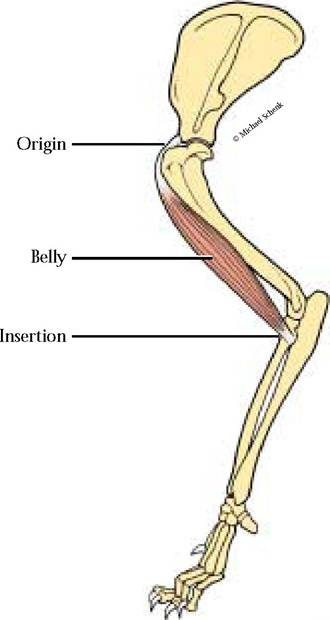
FIGURE 2-1 Gross anatomy of a muscle (Biceps brachii).
and myosin, which are organized into very regular and serially repetitive arrangements. This repetitive banding pattern is a prominent characteristic of striated muscle.
As you will learn during your study of muscles, muscle shapes vary, dependent primarily upon the arrangement of fibers within each muscle and its relationship with the tendon of insertion. Perhaps an arrangement with which you are most familiar is known as convergent [Figure 2-2A]. The fibers in this arrangement are basically parallel; however, they converge at either end of the muscle. The fibers are arranged in parallel throughout the length of strap muscles [Figure 2-2B]. Muscles whose architecture includes a straight blunt origin and a convergent insertion are called fan-shaped [Figure 2-2C]. An oblique arrangement of fibers inserting into a tendon or tendons constitutes the pennate class of muscle architecture. Within this group are unipennate [Figure 2-2D], where the fibers insert into one side of a tendinous insertion, bipennate [Figure 2-2E], where the muscle fibers insert into both sides of a centrally located tendon, and multipennate [Figure 2-2F], where the muscle fibers insert into several tendons whose orientation may vary and may appear as combinations of the unipennate and bipennate subgroups.
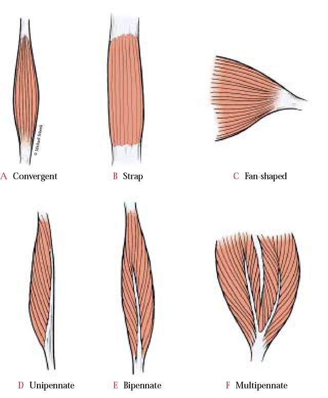
FIGURE 2-2 Muscle architecture.
Muscles are capable of producing a variety of movements called actions. Actions of muscles associated with hinge joints, e.g., the elbow, produce actions known as flexion, causing reduction of the angle at the joint and extension, causing an increase in the angle of the joint.
When appendages or portions of appendages, e.g., the digits, are moved away from a midline reference point or spread, the action is referred to as abduction. In contrast, movement toward the midline reference is called adduction. Movement of an appendage parallel to the longitudinal axis, producing an anterior action, e.g., swinging a leg forward, is known as protraction and the opposite action is known as retraction. Rotation involves the movement of a portion of the body around a central axis, e.g., the head on the neck. A specialized action involving rotation of the radial head in the ulnar notch produces actions known as pronation and supination. When the cat is standing, the manus is pronated or palm down, however, when grooming itself, the manus is supinated or palm up.Individual muscles generally do not bring about actions by themselves. Most actions are the result of the combined effort of several muscles. Those muscles that affect the action directly are called prime movers. Prime movers, however, usually are assisted by others known as synergists. These muscles not only aid in bringing about the main action, but also may stabilize the joint or portions of the skeleton involved in the action and are known as fixators. Muscles whose actions oppose one another are called antagonists.
EXTERNAL FEATURES
Cat specimens are usually packed individually in plastic bags, containing both the cat and some preservative fluid to aid in maintaining a moist environment. Carefully remove the specimen from the bag, retaining the fluid to keep the cat moist when it is returned to the bag for storage.
Lay the cat on its dorsal surface on a large dissecting tray. Now is the time for you to make a number of observations concerning your specimen and also plans for dissection. Note that the body is divided into several regions: a head, neck, trunk, and tail. A number of distinguishing features of the head, all associated with the concentration of special senses in this region, can be seen.
Among them are the paired external ears or pinnae, the paired eyes with an upper eyelid or superior palpebra, and a lower eyelid known as the inferior palpebra, paired nostrils or external nares, and tufts of coarse hairs known as whiskers or vibrissae on either side of the face. Note the nictitating membrane in the lower, medial corner of the eye [Figure 2—3].The trunk can be divided into an anterior thoracic region delineated by the rib cage, a middle abdominal region, and a posterior pelvic region. Along the ventral surface of the trunk, or the belly, are two rows of paired nipples, associated with the mammary glands. They tend to be more prominent in females than in males, especially if the female is either pregnant or has been recently pregnant. Dorsal to the genital region in both sexes and located directly below the tail is the anus, the external terminal opening of the digestive system.
There are two sets of paired appendages, forelimbs, including the manus associated with the cranial portion of the trunk and hindlimbs, including the pes associated with the caudal end of the trunk [Figure 2-3]. Palpate the genital area to ascertain the sex of the cat. If it is a male, you will feel the testes enclosed within the scrotum; if it is a female, note the urogenital aperture.
SKINNING THE CAT
The cat will be skinned on one side only and the skin should be kept in a single piece so that it can be wrapped around the skinned surface when the cat is not being actively worked on. Your instructor may have alternative directions for skinning the cat. Before attempting to remove the skin, observe several possible areas on the body where skin may have been removed to facilitate the injection of blood vessels with latex, e.g., the neck region, the forearm, and the hindleg. Of all the injection sites, the muscles and the blood vessels of the neck region are most likely to be damaged.
Another area that may influence your decision occurs in cats whose hepatic portal system has been injected since the incision in the abdominal area is usually stapled or sutured shut.
In this case, you may want to skin the specimen on the opposite side. If the staples occur in the midventral line, simply choose either side for your incision and cut a flap around the stapled area. Before selection of the side that you wish to skin, observe the position of the injection sites discussed above and choose the pathway that allows you to avoid the majority of problem areas.Since the success of the skinning process is closely correlated with your ability to complete clean, precise cuts, a new blade in your scalpel is essential. Make a careful, shallow incision, just deep enough to break the skin, beginning at the base of the neck 1/2 inch left or right of the midventral line to avoid any muscles whose origin or insertion is on the midline. Determine whether it is possible to pull the skin away from the underlying tissue. Use your fingers, a pair of forceps or a scalpel with the blade held parallel to the underlying muscle or toward the skin and sever the connective tissue from the skin. If your specimen is a female that has been pregnant recently, as you skin the thoracic and abdominal regions you may encounter the mammary glands that will appear as flattened, tannish masses that you might mistake for muscle. It is preferable to remove these glands with the skin. Continue caudally to a level approximately two inches anterior to the cranial edge of the hindlimb. Now angle your incision along the midline of the hindlimb continuing to a point just proximal to the digits where you will make an encircling incision around the pes. If your specimen is a male, exercise extreme caution because the spermatic cord is imbedded within the fat and connective tissue of the
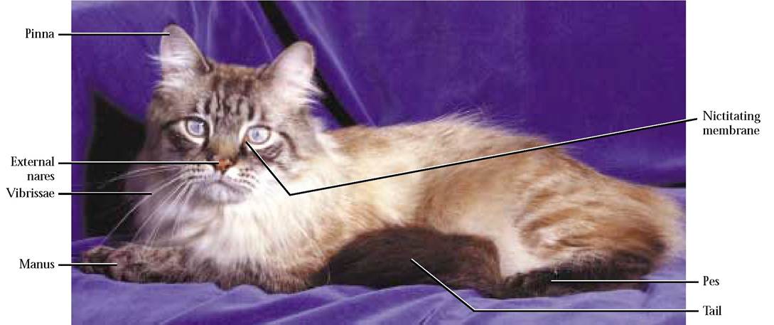
FIGURE 2-3 External features of a cat. (This cat was not used as a dissection specimen in this book.)
groin area and directly beneath the skin. Another reason for As you loosen the skin from the underlying tissues, make a
this care is that the leg skin is very thin and a major super- circular incision around the ear and reflect the skin.
Con-ficial vein, the saphenous, lies directly under the skin. There- tinue the skinning process to approximately 1/2 inch past
fore, carefully sever the fascia from the skin in this area.
Return to the thoracic region and begin an incision opposite the forelimb, continuing down the medial aspect, and encircling the manus just proximal to the digits. Be exceptionally cautious when skinning the radial side of the lower forelimb since a thin, narrow muscle band, the brachioradialis m., a nerve, and blood vessels, adhere closely to the skin and may be mistakenly severed. In addition, along the lateral aspect of the forelimb, from the wrist to the shoulder, courses the cephalic vein, that can again, very easily, be removed along with the skin. It should not be, however! Carefully skin the body, hindlimb and forelimb. As the trunk is skinned, an extensive dermal muscle, the cutaneous maximus m., especially prominent in the axillary, pectoral, and abdominal regions, will be encountered. This is the muscle in horses that allows them to twitch and get rid of flies, dogs to shake water out of their coats, etc. It is best to remove this muscle with
the mid-dorsal line along the entire length of the cat [Figure 2-4A, Figure 2-4B, and Figure 2-4C].
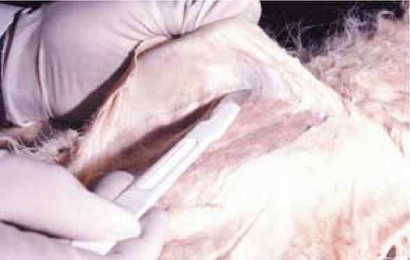
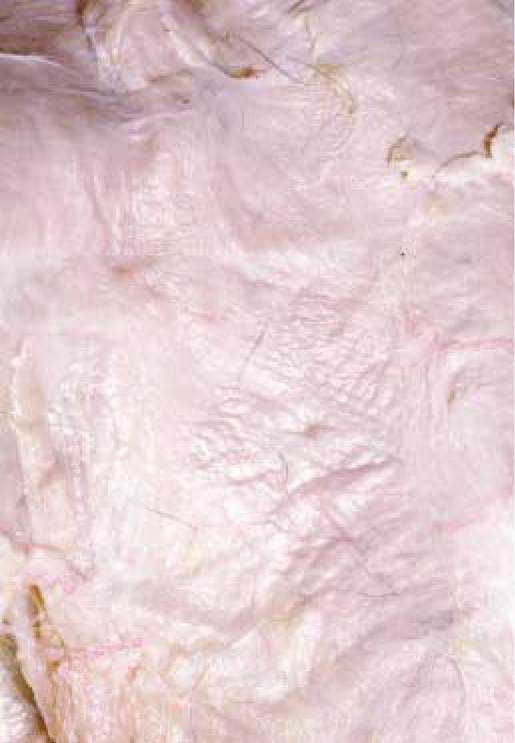
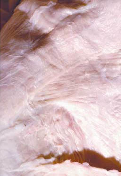
A Proper skinning technique. The edge of the scalpel blade is held next to the skin.
the skin, exerting great care in the axillary region. In the dorsal shoulder region, take care not to cut through a heavy white connective tissue (aponeurosis) between the paired acromio- trapezius muscles.
Concentrate now on the neck and head regions. Be careful of
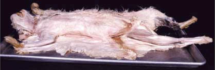
B Cat properly skinned.
superficial blood vessels in the ventrolateral position in the neck. During your dissection of this area, note another dermal muscle, the platysma m., that adheres closely to the neck and head muscles. Again, it is desirable to remove the platysma with the skin. Notice that the skin in the head region is much thicker than other areas of the body. Extend the ventral incision to the base of the mandible, outlining the mouth, the nose, and the eye, continuing the incision to the midline of the forehead.
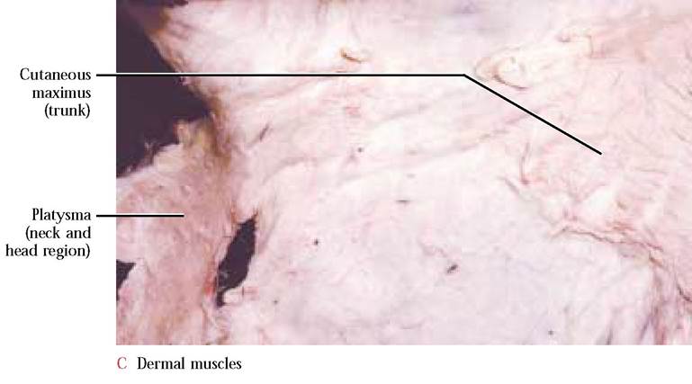
FIGURE 2-4 Skinning the cat.
Humans lack a cutaneous maximus and, for this reason, they do not have the ability to twitch their skin, and scar much more easily than other mammals. The platysma, however, is well developed in humans. As you look in a mirror, grimace and note the tendinous, stringy appearance of your neck, the action of the platysma.
Preparing the cat for
MUSCLE DISSECTION
To properly separate and appreciate muscle relationships, it is necessary to remove extraneous tissues that tend to adhere to the surface of the superficial muscles [Figure 2-5A and Figure 2-5B]. Carefully remove fatty tissue lying on external muscle surfaces. Usually there is a very heavy deposit of fat in the groin area. If your specimen is a male, exercise extreme caution during the removal of fat in this region since the spermatic cord, a thin, small diameter tube lies in close proximity to this fat deposit. Another area where fat may accumulate is the region between the scapulae on the surface of the aponeurosis connecting the two acromio- trapezius muscles. Exert caution while removing this fatty tissue here so that the aponeurosis is not destroyed. There will be other areas on the body surface where heavy sheets of dense connective tissue covered by fatty tissue may occur (lumbosacral region, the insertion end of the tensor fascia latae muscle, biceps femoris muscle, etc.). Caution should always be exerted to avoid damaging these important areas of fascia and any other underlying structures. Fascia associated with muscle insertions should not be removed.
After skinning the cat, there may be pieces of the dermal muscles (platysma and cutaneus maximus) left adhering to the muscle surfaces. These should be carefully removed. The epitrochlearis muscle appears very much like a piece of cutaneus maximus on the medial surface of the brachium [Figure 2-27A]. Do not remove this muscle or the thin aponeurosis by which it inserts in the vicinity of the elbow.
A Before removal of overlying connective tissue.
B After removal of overlying connective tissue showing an example of differences in muscle fiber direction in contiguous or overlying muscles.
DIRECTION
OF MUSCLE FIBERS
During the dissection of your specimen, it is very advantageous to be able to distinguish where one muscle ends and an adjacent or overlapping muscle begins. In order to identify individual muscles, look for the direction of muscle fiber orientation. For example, in muscles such as the abdominals, consisting of three sheetlike layers superimposed one on the other, it becomes essential to detect changes in fiber direction [Figure 2-10B]. Furthermore, most muscles are individually wrapped in layers of connective tissue called fascia and the areas where these layers abut one another can often be observed as distinct lines between muscles [Figure 2-5A, Figure 2-5B]. In order to separate muscles that overlap or abut one another, it I is imperative to orient the probe or.
fingers within the fascia separating those muscles to ensure that one is not creating new muscles. There is nothing more frustrating than contending with several muscles where only one should exist. Your ability to distinguish and separate contiguous muscles will be greatly enhanced by training yourself to appreciate these relationships and will be greatly appreciated by your instructor.
SEWING AND CUTTING MUSCLES
Sewing a muscle may sound strange to you, but contending with the cut ends of several chest or leg muscles might change your mind. The sewing and cutting process maintains the integrity of the points of origin and insertion of the muscle. Since you will be identifying superficial and deep muscles, our solution is to first sew and cut the more superficial muscles and then separate the underlying muscles.
Before attempting to sew and cut any muscle, separate the muscle from the point of origin to the point of insertion and free it from contiguous muscles. You are now ready to sew the muscle:
1. With approximately 18 inches of thread, thread the needle leaving one end longer than the other.
2. Make a knot only in the longer of the two free ends.
3. Insert the needle approximately 1/2 inch from the midline between the origin and insertion of the muscle, pull it through and make a couple of overcast stitches to thoroughly anchor the thread [Figure 2-6A]. Since the muscle will always be cut perpendicular to the muscle fibers, orient your sewing points accordingly.
4. Insert the needle approximately 1/2 inch from the midline of the muscle on the other side. Notice that there will be about an inch of space between the two anchor points [Figure 2-6B].
5. Pull the thread through, leaving a loop approximately three inches in length [Figure 2-6B].
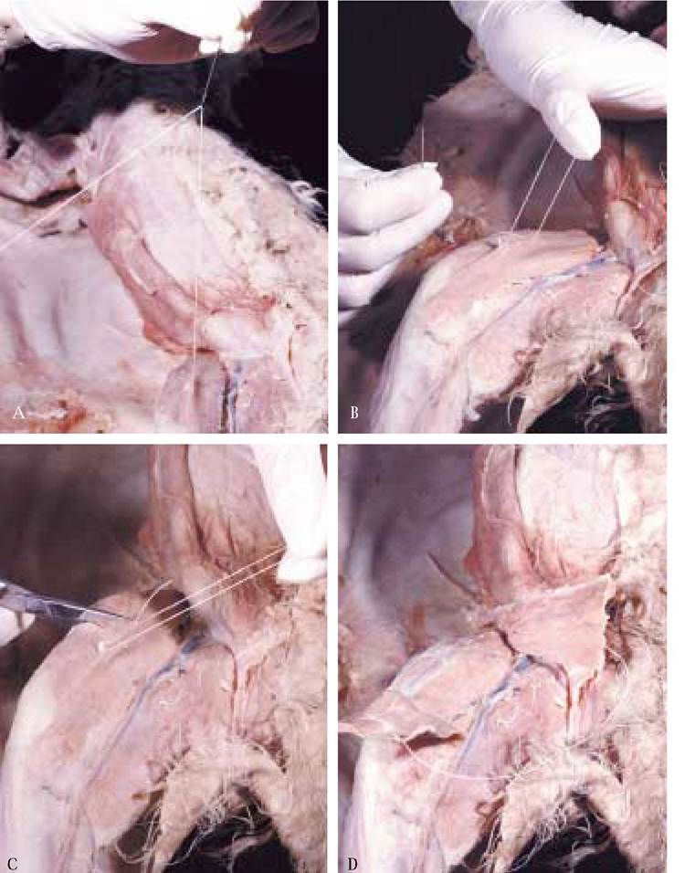
FIGURE 2-6 How to sew and cut a designated muscle.
6. Similar to the other side, make two to three overcast stitches to securely anchor the thread.
7. Cut the thread off at the second anchoring site, leaving the loop attached to the muscle.
8. Lift the muscle and carefully cut through the midline of the muscle only, leaving the loop of thread connecting the two ends [Figure 2-6C and Figure 2-6D]. Remember to cut each muscle perpendicular to the muscle fibers.
Pectoantebrachialis m.
This is the most superficial of the chest or pectoral muscles. It is a narrow, thin band that extends from the midline of the body to the upper portion of the forelimb [Figure 2-7]. This muscle does not occur in man.
Origin: Manubrium of the sternum
Insertion: Flat tendon into the superficial fascia of the antebrachium above the elbow
Action: Draws the forelimb toward the midline
SUPERFICIAL
THORACIC MUSCLES
**Sew and cut this muscle.
This group of muscles has a tendency to adhere tightly to one another, therefore, care should be exercised when separating them. Watch for the changes in muscle fiber orientation and the subtle white lines created by the connective tissue surrounding each muscle that indicate the extent of individual muscles.
Insertion: Middle third of the shaft of the humerus
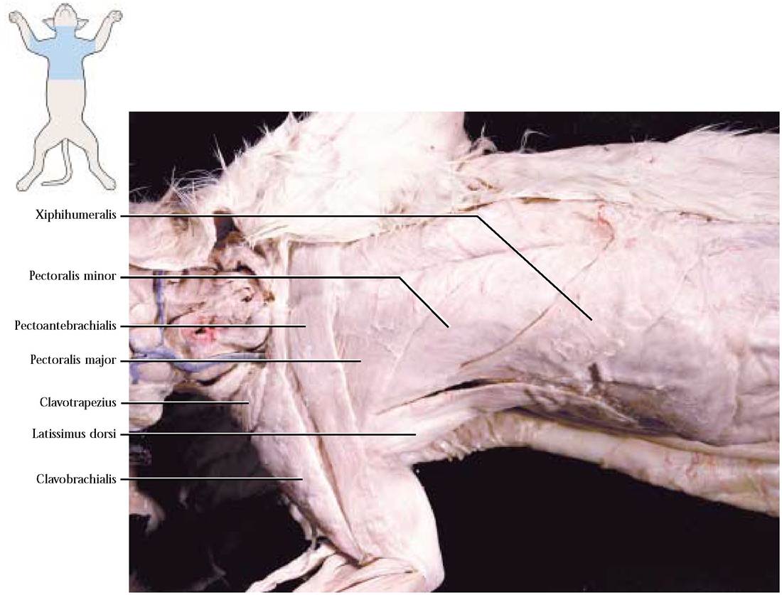
Pectoralis major m.
A superficial and deep portion of this muscle can be distinguished [Figure 2-7, Figure 2-8A, and Figure 2-8B].
Superficial Portion
Flat, thin band, approximately the same width as the pectoantebrachialis and partially hidden by that muscle [Figure 2-8A].
Origin: Midventral raphe and cranial half of the manubrium

FIGURE 2-8 Pectoralis major.
Deep Portion
Flat band, approximately three times the width of the superficial part [Figure 2-8B]. In order to see the entire extent of this portion, the Clavotrapezius and the clavo- brachialis should now be dissected (see pages 52 and 53). Exercise care while separating the clavotrapezius to avoid damaging the underlying pectoralis major. Furthermore, use care in separating this portion of the pectoralis major to avoid damaging the underlying pectoralis minor.
Origin: Cranial half of the sternum and midventral raphe Insertion: Proximal third of the shaft of the humerus Common Action of Both Portions: Draws the forelimb toward the midline and turns the manus forward
**Sew together and cut both portions of this muscle following careful separation of the two portions.
Pectoralis minor m.
A thick, fan shaped muscle extending caudally to and beneath the deep portion of the pectoralis major [Figure 2-7 and Figure 2-9]. Exert care to preserve the xiphihumeralis that passes beneath the pectoralis minor [Figure 2-7, Figure 2-8A, and Figure 2-8B]. Additionally, with great care, separate the latissimus dorsi from the lateral border of the pectoralis minor [Figure 2-7].
Origin: From the six sternebrae and sometimes the xiphoid process, resulting in the appearance of several slips that appear to be separate muscles
Insertion: Ventral border of the humerus from the bicipital groove to the middle of the humerus
Action: Draws the forelimb toward the midline
**Sew and cut this muscle.
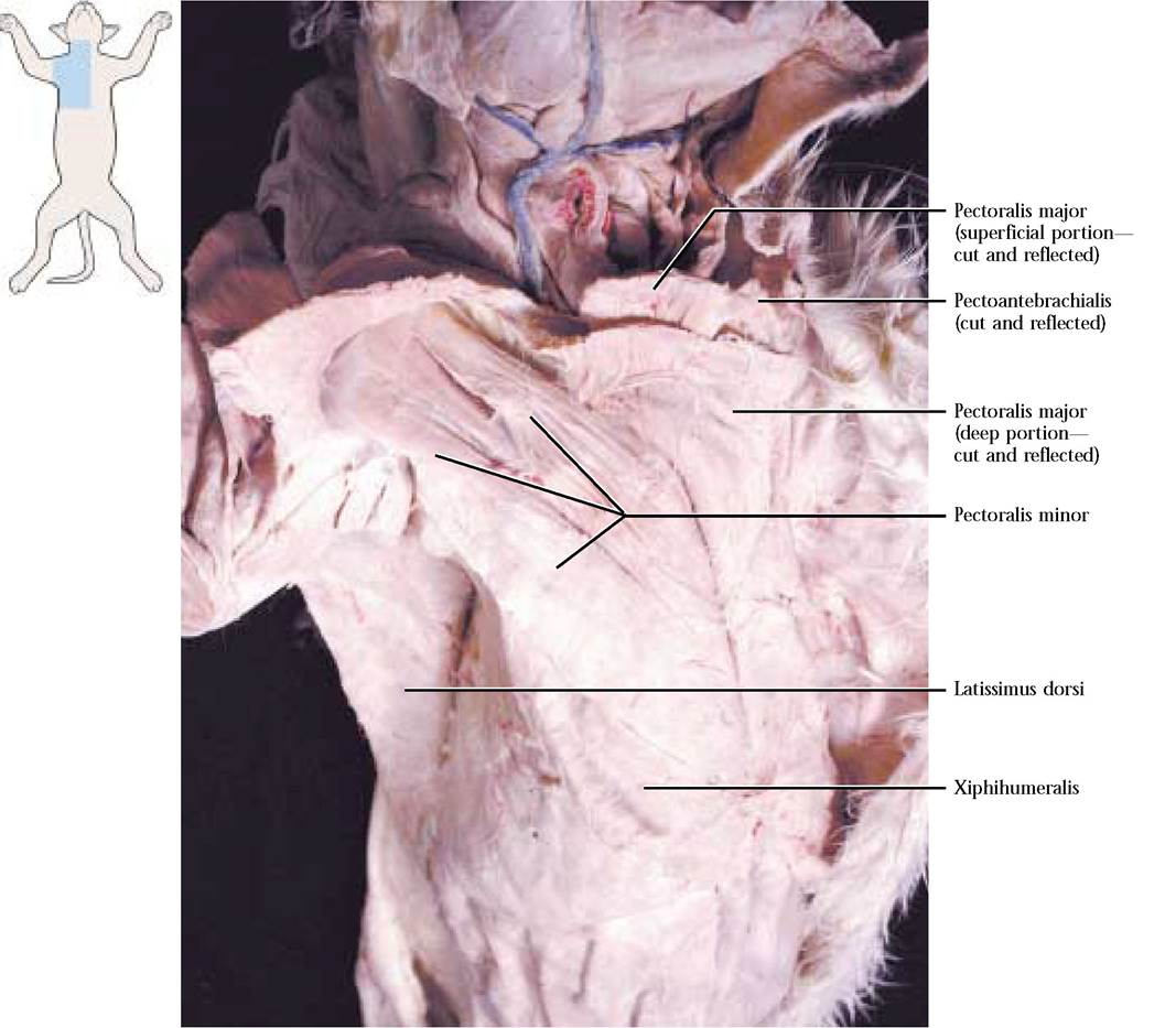
FIGURE 2-9 Pectoralis minor.
Xiphihumeralis m.
A long, very thin, narrow band of muscle, lying along the posterior border of the pectoralis minor and, according to some anatomists, actually a part of that muscle [Figure 2-7 and Figure 2-9]. The Xiphihumeralis muscle is absent in humans.
Origin: Median raphe in the vicinity of the xiphoid process
Insertion: Along the ventral border of the bicipital groove of the humerus
Action: Synergistic with the pectoralis minor in drawing the forelimb toward the midline
**Sew and cut this muscle.
ABDOMINAL MUSCLES
Three sheetlike muscles and a longitudinal, bandlike muscle make up this group. Note that the left and right portions of the abdominal muscles are separated by a longitudinal white line of connective tissue known as the linea alba [Figure 2- 10A]. The sheetlike muscles are thin and quite extensive, supporting the entire abdominal area and a portion of the ventral thoracic region. These muscles are layered and adhere closely to one another by means of fascia. The direction of fibers within each sheet is distinctive and this feature is used as a tool to identify the individual muscles. To facilitate the dissection of these sheets, a three sided opening, one inch on each side should be made in the flank
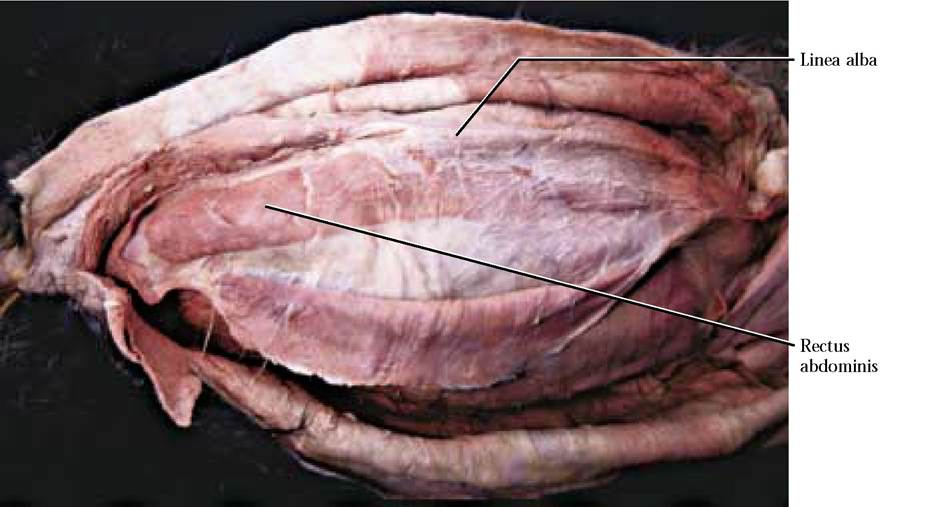
A Linea alba and rectus abdominis
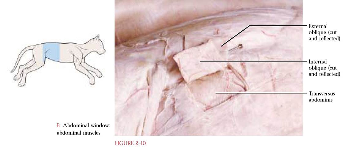
[Figure 2-10B]. Carefully separate and identify the sheets of muscle.
External oblique m.
The direction of the fibers of this muscle extend cranio- dorsally [Figure 2-10B]. This is the most superficial of the three sheetlike abdominal muscles.
Origin: Lumbodorsal fascia and the last 9 or 10 ribs
Insertion: Median raphe of distal portion of sternum, linea alba from sternum to pubis
Action: Compresses the abdominal region
Internal oblique m.
The direction of the fibers of this muscle extend caudo- dorsally [Figure 2-10B]. This sheetlike muscle lies directly beneath the external oblique.
Origin: Lumbodorsal fascia in common with the external oblique and iliac crest
Insertion: Linea alba by a thin aponeurosis in common with the external oblique and transversus abdominis
Action: Compresses the abdominal region
Transversus abdominis m.
Fibers of this muscle sheet extend nearly transversely between the origin and insertion [Figure 2-10B]. This muscle sheet lies directly beneath the internal oblique.
Origin: Aponeurosis from the costal cartilages of the vertebrochondral and vertebral ribs, transverse processes of lumbar vertebrae and ventral border of the ilium
Insertion: Linea alba in common with the two obliques
Action: Compresses the abdomen
Rectus abdominis m.
This muscle occurs as a longitudinally directed band of muscle on either side of the linea alba [Figure 2-10A]. This muscle is encased in a sheath formed by the aponeuroses of the other three abdominal muscles. In humans, this muscle is reduced at its anterior end.
Origin: Tubercle of pubis
Insertion: First and second costal cartilage, proximal end of sternum by a tendon passing dorsal to the transversus costarum
Action: Compresses the abdominal region, pulls sternum and ribs caudally causing flexion of the trunk.
SUPERFICIAL
BACK MUSCLES
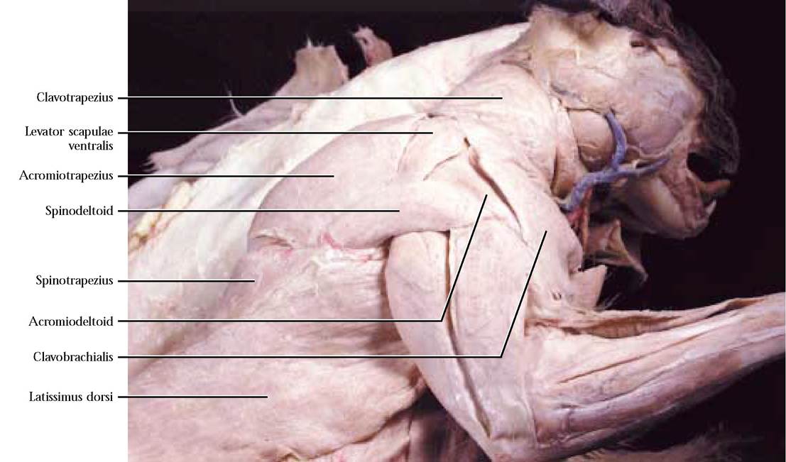
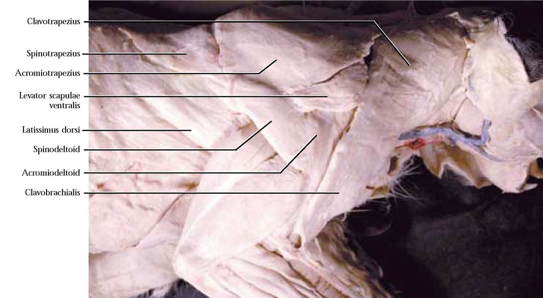
Clavotrapezius m.
This is a wide, flat muscle that covers most of the lateral portion of the neck [Figure 2-11 and Figure 2-12]. Take note that the levator scapulae ventralis passes below the clavotrapezius and must be separated from the clavo- trapezius before the clavotrapezius is cut.
Origin: Lambdoidal ridge, middorsal raphe over spine of the axis
Insertion: Clavicle and raphe between clavotrapezius and clavobrachialis
Action: Protraction of the humerus
**Sew and cut this muscle.
Clavobrachialis m.
This muscle appears to be a continuation of the clavo- trapezius onto the forelimb and is considered by some anatomists to be the cranial portion of the deltoid and is called the clavodeltoid [Figure 2-13 and Figure 2-14]. The clavotrapezius and the clavobrachialis are separate muscles.
Origin: Clavicle and raphe between Clavotrapezius and Clavobrachialis
Insertion: Commonly inserted with the brachialis through a tendon on the medial surface of the ulna distal to the semilunar notch
Action: Flexes the forearm
**Sew and cut this muscle.
Acromiotrapezius m.
This is a thin trapezoidal muscle lying over the scapulae [Figure 2-13 and Figure 2—14]. Extreme care must be exercised while dissecting this muscle to prevent damage to the whitish middorsal tendon and fascia that hold the left and right acromiotrapezius muscles together over the vertebral column.
Origin: Middorsal line from the spine of the axis to the spinous process of the fourth thoracic vertebra
Insertion: Metacromion process and spine of the scapula Action: Adduct and stabilize the position of the scapulae
**Sew and cut this muscle, not the aponeurosis!
Spinotrapezius m.
This is a triangular muscle and the most posterior of the trapezius group [Figure 2—13 and Figure 2—14]. With great care, dissect this muscle from the craniodorsal surface of the latissimus dorsi.
Origin: Originates from the spinous processes of most of the thoracic vertebrae
Insertion: Fascia of supraspinatus and infraspinatus muscles on either side of the spine
Action: Pulls the scapula dorsally and caudally
The human trapezius is represented by a fusion of the three trapezius muscles in the cat.
**Sew and cut this muscle.
Latissimus dorsi m.
This is a large, thick, flat, triangular muscle just posterior to the trapezius group and covered craniodorsally by the spinotrapezius [Figure 2-11 and Figure 2-12]. The reason that the axillary region often appears to be so ragged is that the cutaneus maximus has its cranial origin in the axilla and has probably been cut off and left attached to the skin.
Origin: Neural spines of the fourth or fifth thoracic to the sixth lumbar vertebrae
Insertion: Medial surface of shaft of humerus at the proximal end
Action: Pulls forelimb dorsocaudally
**Sew and cut this muscle.
DEEP THORACIC MUSCLES
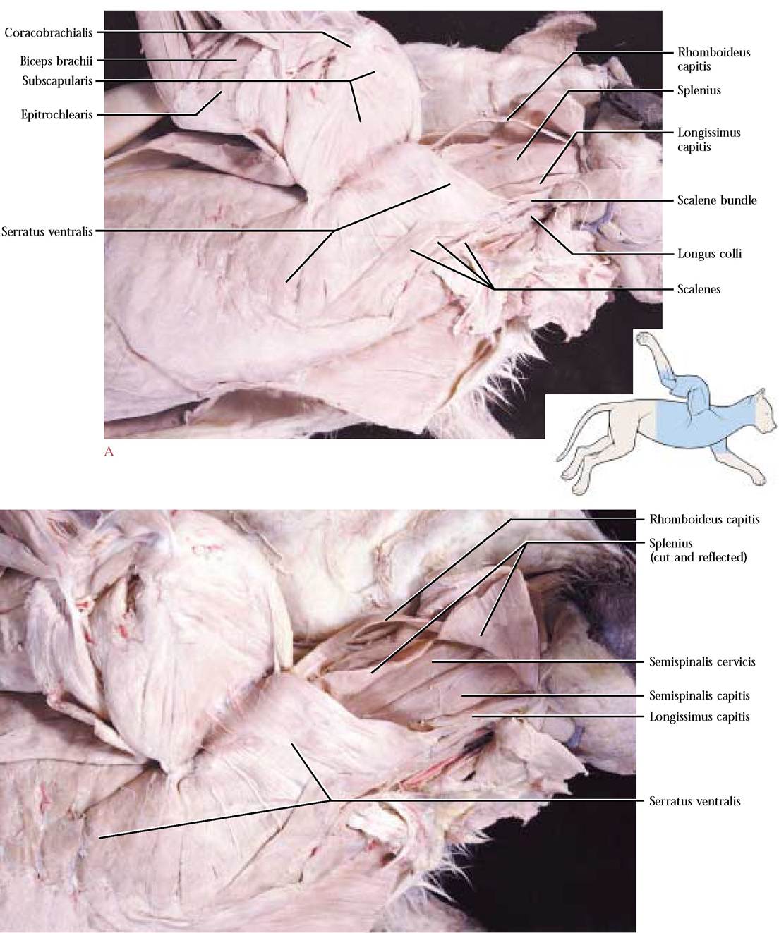
Serratus ventralis m.
This is a large, fan-shaped muscle, made up of obvious individual slips, extending between the thorax and the scapula [Figure 2-13]. Notice that these individual slips are more conspicuous at the caudal end of this muscle. In the human, this muscle is represented by two separate muscles, a cranial levator scapulae originating from the cervical vertebrae and the caudal serratus anterior originating from the ribs.
Origin: From the surface of the first nine or ten ribs and the transverse processes of the last five cervical vertebrae
Insertion: Vertebral border of the scapula
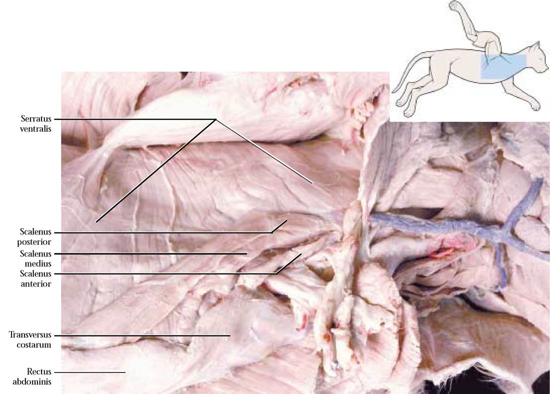
FIGURE 2-13 Cranial deep thoracic muscles. NOTE: This cat had an unusually long scalenus posterior.
Action: Draws the scapula toward the thoracic wall and helps to support the scapula
Scalenus anterior, posterior, and medius m.
These three bandlike muscles, scalenus anterior, the most ventral, scalenus posterior, the most dorsal, and scalenus medius, situated between the two, lie at an oblique angle along the lateral aspect of the thorax [Figure 2—13]. Cra- nially, these three muscles unite into a single band or bundle known as the scalene bundle [Figure 2-20A].
Origin: S. anterior m.—from the second and third ribs, S. posterior m.—from the third or fourth ribs, S. medius m.—from the sixth through the ninth ribs
Insertion: Transverse processes of all cervical vertebrae
Action: Bends the neck and pulls ribs cranially
** This is an example of a muscle whose origin and insertion may be reversed, thereby producing contrasting actions depending upon the fixed end.
Transversus costarum m.
This is a thin, bandlike muscle extending from the sternum and covering the cranial portion of the rectus abdominus muscle [Figure 2—13].
Origin: From the side of the sternum
Insertion: First rib and costal cartilage
Action: Pulls the ribs cranially
Intercostalis externus m.
Note that the fibers of this outer layer of muscles lying in the intercostal spaces between adjacent ribs are oriented craniodorsally, similar to the external oblique layer, from which they were derived [Figure 2—14]. These muscles are absent from the costal cartilages of the first through the seventh ribs.
Origin: From a cranial rib
Insertion: To an adjacent caudal rib
Action: Protracts the ribs
Intercostalis internus m.
This layer lies directly below the external intercostals [Figure 2-14]. With a sharp scalpel and a light touch, make a threesided ½ inch window and carefully peel the intercostalis externus layer back. Note that the fibers of this muscle are oriented caudodorsally, similar to the internal oblique layer, from which they were derived.
Origin: From a caudal rib
Insertion: To an adjacent cranial rib
Action: Retracts the ribs
Transversus thoracis m.
This incomplete third layer lies beneath part of the internal intercostals and represents the thoracic portion of the transversus abdominis. It can best be seen later during the dissection of the thoracic cavity [Figure 5-4].
Origin: Dorsolateral border of the sternum between the third to the eighth rib
Insertion: Costal cartilages near the junction of the ribs
Action: Moves ribs
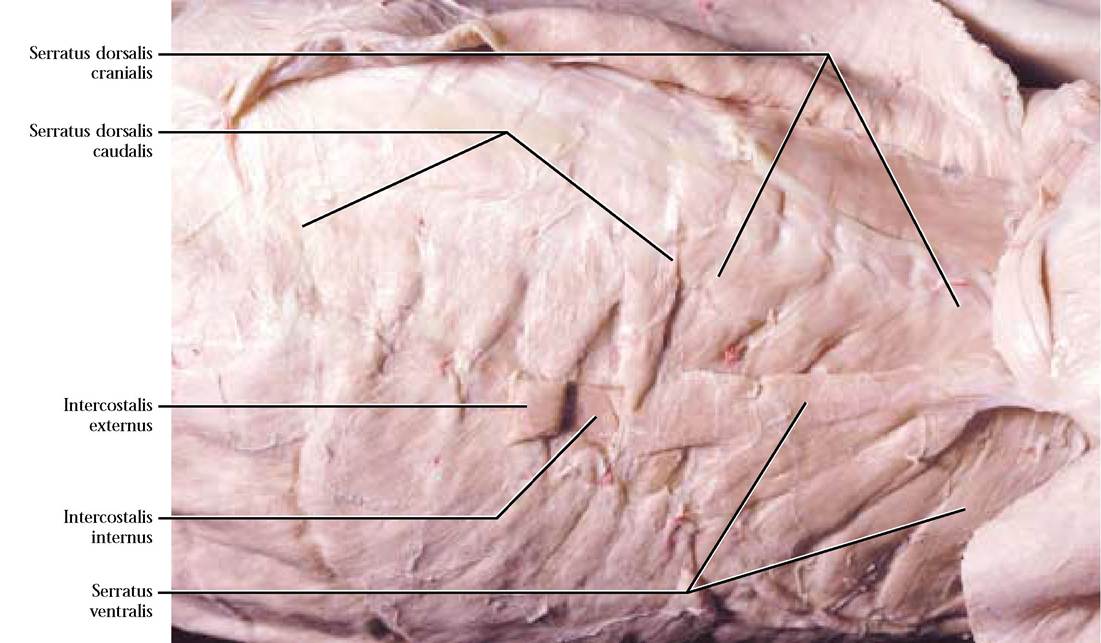
Serratus dorsalis cranialis m.
This thin layer of muscle appearing as slips extends along the dorsal part of the thorax and neck beneath the latissimus dorsi [Figure 2—14].
Origin: From the cervicothoracic middorsal fascia
Insertion: On the outer surface of the first nine ribs
Action: Draws the ribs cranially
Serratus dorsalis caudalis m.
This thin layer of muscles, also appearing as slips, extends from the caudal end of serratus dorsalis cranialis to the lumbar region [Figure 2-14]. Note that, although appearing continuous, serratus dorsalis cranialis and serratus dorsalis caudalis possess fibers that are oriented in directions opposite one another.
Origin: From the lumbar middorsal fascia
Insertion: Last four or five ribs
LOWER BACK MUSCLES
To begin the dissection of the lower back and thoracic muscles use a pair of forceps to hold up the lumbodorsal fascia, make a small hole in the double layered fascia and then cut a three-sided window approximately two inches on a side [Figure 2—15]. Depending upon the size of the cat, the dimensions of the window may vary.
LUMBAR AND THORACIC
Multifidus spinae m.
This is an extensive muscle consisting of many bundles of fibers that can best be distinguished abutting the vertebral column in the lumbar region [Figure 2-15].
Origin: Primarily transverse processes of vertebrae
Insertion: Neural spines of more anterior vertebrae
Action: Draws the ribs caudally
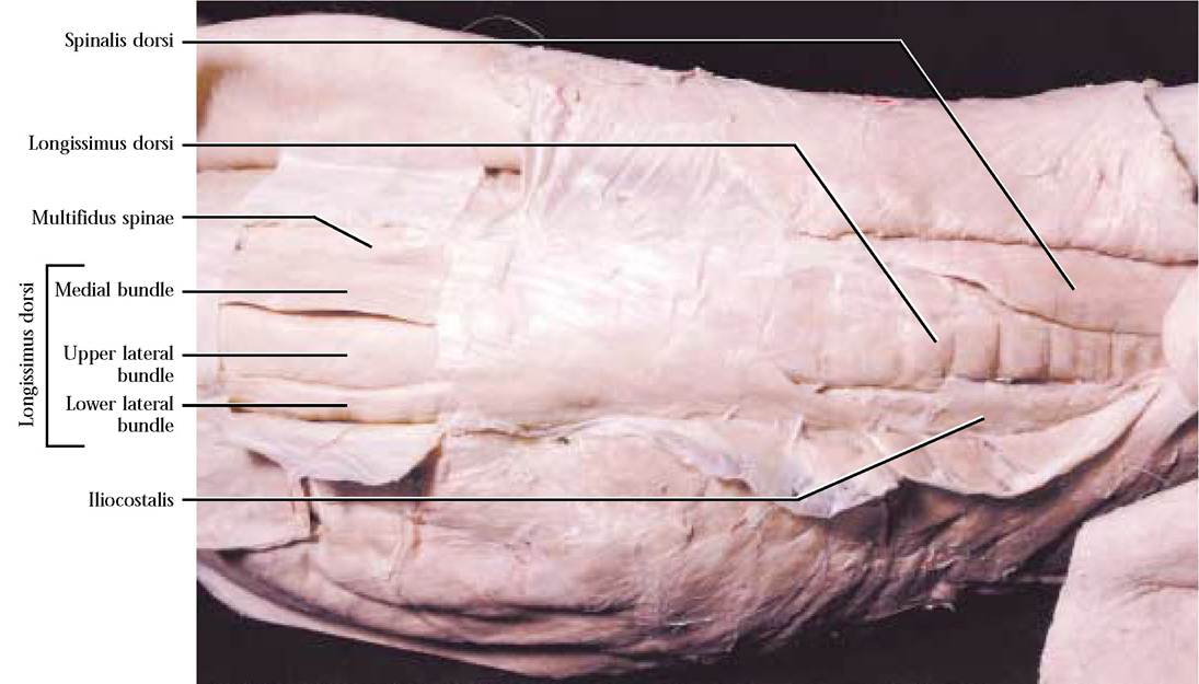
FIGURE 2-15 Caudal back window: lumbar and thoracic back muscle complex.
Action: When both sides contract simultaneously, extends the vertebral column; when contracted unilaterally, bends the vertebral column toward that side
Longissimus dorsi m.
This is another extensive muscle occupying the space between the neural spines and transverse processes and extending from the prominent lumbar region to the much less bulky thoracic and cervical regions [Figure 2—15]. There are a number of distinguishable bundles in the lumbar region, a medial and a lateral that is further subdivided by fascia into upper and lower lateral portions. Note that the longissimus capitis is the cervical extension of the longissimus dorsi.
Origin: Medial bundle—From neural spines of vertebrae in the vertebral column; Lateral bundle—From the ilium and neural spines of the vertebrae in the vertebral column
Insertion: Onto various processes of more anterior vertebrae
Action: Extends the vertebral column
Spinalis dorsi m.
This muscle is formed as a medial separation of the longissimus dorsi in the thoracic region [Figure 2—15].
Origin: From the neural spines of more
posterior thoracic vertebrae
Insertion: Transverse processes of more cranial vertebrae
Action: Extends the vertebral column
Iliocostalis m.
This is a thin muscle confined to the thoracic region consisting of a number of bundles lying lateral to the longissimus dorsi over the dorsal aspect of the ribs [Figure 2-15].
Origin: Lateral surface of the ribs
Insertion: Onto the lateral surface of more cranial ribs
Action: Pulls the ribs together
MUSCLES OF THE NECK
Sternomastoid m.
The origin of this paired, bandlike muscle forms the apex of a V just cranial to the anterior end of the sternum while the arms of the V continue toward the base of the ear [Figure 2-16A and Figure 2-16B].
Origin: Cranial end of the manubrium
Insertion: Lambdoidal ridge and mastoid portion of the temporal bone
Action: As a pair—flexion of the head
Individually—turns the head
Cleidomastoid m.
A flat, bandlike muscle that extends between the clavicle and the temporal region of the skull lies dorsolateral to the sternomastoid [Figure 2-16B].
Origin: Mastoid process of the temporal bone
Insertion: Clavicle
Action: When clavicle is stationary—turns the head; When head is stationary—moves the clavicle anteriorly
**In the human, the sternomastoid and the cleidomastoid muscles are united into a single muscle, the sternocleidomastoid with similar actions.
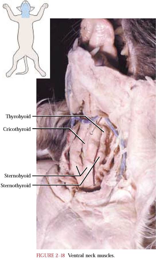
Sternohyoid m.
A slender, bandlike muscle that lies along either side of the midventral line of the neck [Figure 2-17].
Origin: First costal cartilage
Insertion: Hyoid bone
Action: Retracts the hyoid
Sternothyroid m.
This slender, bandlike muscle lies somewhat dorsal to the sternohyoid and lateral to the trachea [Figure 2-18].
Origin: First costal cartilage
Insertion: Thyroid cartilage of the larynx
Action: Retracts the larynx
Thyrohyoid m.
This is a small, bandlike muscle lying along the lateral aspect of the larynx [Figure 2-18].
Origin: Lateral portion of the thyroid cartilage of the larynx
Insertion: Hyoid bone
Action: Protracts the larynx
Cricothyroid m.
This broad, short, flat muscle band lies on the ventral surface of the cricoid cartilage of the larynx [Figure 2-18].
Origin: Surface of the cricoid cartilage
Insertion: Thyroid cartilage
Action: Regulates tension of the vocal cords
Stylohyoid m.
Mylohyoid m.
This is a very slender band stretching across the posterior surface of the digastric m. [Figure 2-16A]. Great care must be exercised in exposing this small, slender muscle because it is overlain with connective tissue making it especially vulnerable to removal.
Origin: Stylohyal, a segment of the lesser cornua of the hyoid
Insertion: Body of the hyoid
Action: Elevates the hyoid
Digastric m.
This thick muscle lies along the medial ventral border of the mandible [Figure 2-16A].
Origin: From the mastoid and jugular processes
Insertion: Medial ventral border of
the mandible
Action: Depresses the mandible
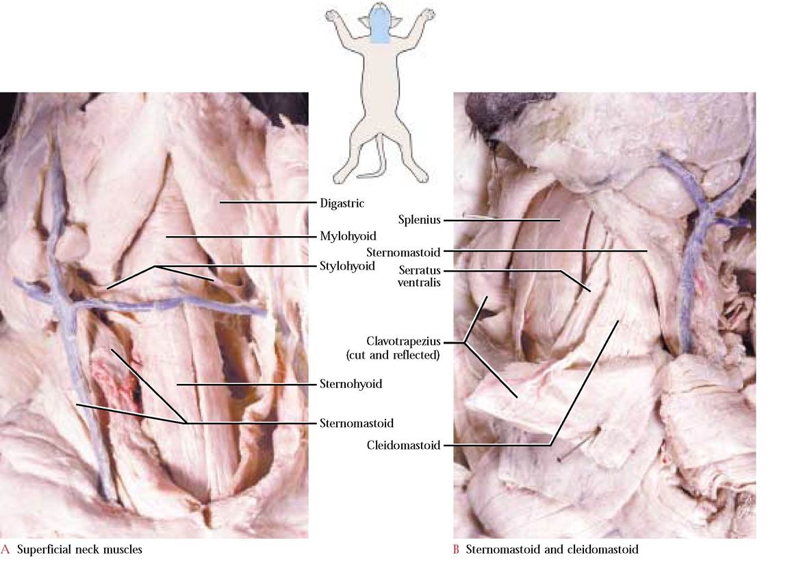
This thin, roughly triangular muscle with distinct transverse fibers, lying between the two dentary bones of the mandible, consists of a pair of muscles connected by a thin, tendinous median raphe [Figure 2-16A]. The raphe extends from the mandibular symphysis to the hyoid. With a sharp scalpel make an incision through the median raphe. Carefully reflect one of these thin muscles toward the mandible, taking care not to shred its fibers.
Origin: Medial surface of the mandibular body Insertion: Median raphe
Action: Elevates the floor of the mouth
Geniohyoid m.
A narrow, elongated muscle that lies along the median raphe dorsal to the mylohyoid [Figure 2-17].
Origin: Ventral surface of the mandible just lateral to the symphysis
Insertion: Body of the hyoid
Action: Protracts the hyoid
Genioglossus m.
This is another narrow, elongated muscle that lies dorsolateral to the geniohyoid [Figure 2-17].
Origin: Ventral surface of the mandible, near the symphysis, and dorsal to the geniohyoid
Insertion: Tongue
Action: Draws tip of the tongue backward and the root forward
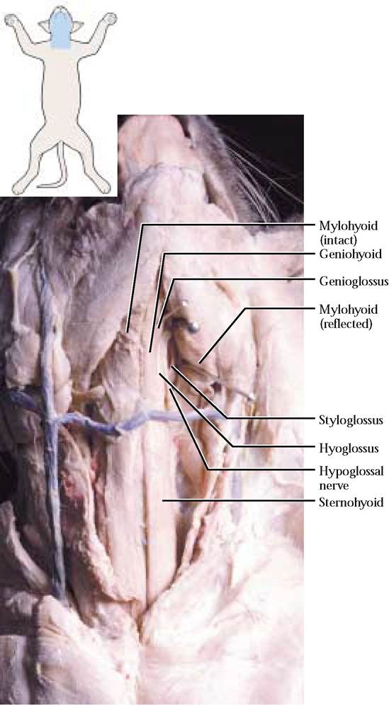
Hyoglossus m.
Lateral to the geniohyoid lies this roughly rhomboidal muscle [Figure 2-17]. It can be readily identified since the hypoglossal nerve (C.N. XII) lies over its surface.
Origin: Body of the hyoid
Insertion: Tongue
Action: Retracts and depresses the tongue
Styloglossus m.
This is a bandlike muscle lying lateral to the hyoglossus and parallel to the digastric m. [Figure 2-17]. To observe the styloglossus, carefully pull the digastric muscle laterally.
Origin: Mastoid process
Insertion: Tongue
Action: Retracts and elevates the tongue
Intrinsic muscles of the tongue are entirely contained within the body of the tongue and are known as the lingualis proprius. They make up the bulk of the tongue and assist in tongue movement.
Origin: Caudal portion of the cervical supraspinous ligament and vertebral spines of the first four thoracic vertebrae
Insertion: Vertebral border and outer surface of the scapula
Action: Adducts scapulae
DEEP NECK AND BACK MUSCLES
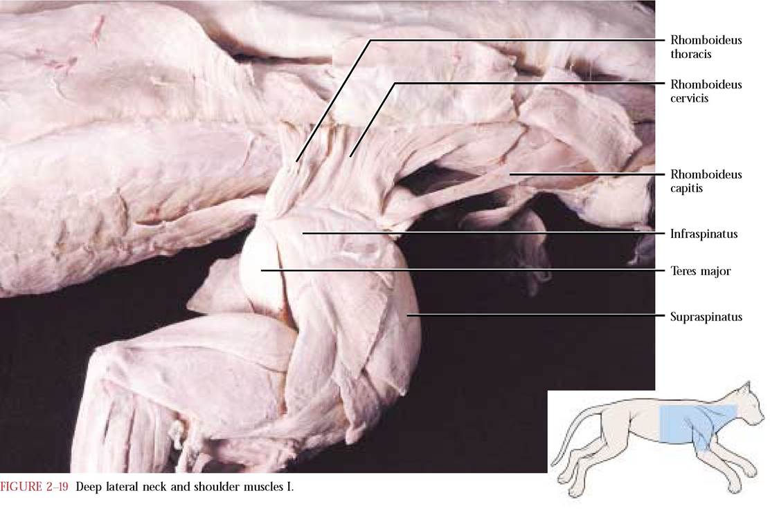
Rhomboideus capitis m.
This is the cranial portion of the rhomboideus complex that consists of a narrow, thin, flat, lateral band [Figure 2—19].
Origin: The mediolateral half of the lambdoidal ridge
Insertion: Along the dorsal border of the scapula
Action: Rotates and pulls scapula cranially
Rhomboideus cervicis and thoracis m.
This portion of the complex is a thick trapezoidal muscle separated into an anterior cervical and posterior thoracic part [Figure 2-19].
Splenius m.
The fibers of the splenius and longissimus capitis muscles often adhere to one another. Look for the subtle white line of connective tissue holding these two muscles together. With a pair of fine forceps carefully separate the two.
The splenius, a large, flat muscle on the dorsolateral aspect of the neck lies beneath the rhomboideus capitis [Figure 2-20A].
Origin: Midorsal line of neck and adjacent fascia Insertion: Along the lambdoidal ridge of the occipital bone
Action: The joint action of the left and right splenius muscles elevate or extend the head, individually, each muscle flexes the head laterally
B
FIGURE 2-20 Deep lateral neck and shoulder muscles II.
Longissimus capitis m.
This narrow, straplike muscle, a cranial continuation of the longissimus dorsi, lies along the ventral edge of the splenius [Figure 2-20A and Figure 2-20B].
Origin: By several tendons from the prezygapophyses of cervical vertebrae 4-7
Insertion: Mastoid process of the temporal bone
Action: Lateral flexion of the head
The next two muscles are sometimes identified as the semispinalis cervicis and capitis. These are flat muscles lying beneath the splenius [Figure 2-20B]. In order to identify these muscles, it will be necessary to bisect the splenius at right angles to its fibers and reflect the halves.
Semispinalis cervicis m.
Origin: Spinous processes of cervical vertebra 7 and the first three thoracic vertebrae
Insertion: Median third of the lambdoidal crest
Action: Elevates the head
Semispinalis capitis m.
Origin: Prezygapophyses of cervical vertebrae 3-7 and first three thoracic vertebrae
Insertion: Median third of the lambdoidal crest
Action: Elevates the head
Longus colli m.
This is a narrow band of muscle that lies along the lateral aspect of the neck ventral to the scalene bundle [Figure 2-20A].
Origin: From the ventral surfaces of the first six thoracic vertebrae, and ventral surfaces of bodies and transverse processes of the cervical vertebrae
Insertion: Slips from the thoracic vertebrae unite and insert commonly onto the ventral portion of the sixth cervical transverse process, while slips from the cervicals extend cranially to insert on the midline of the centra of more anterior cervical vertebrae
Action: Bends the neck both ventrally and laterally
MUSCLES OF THE HEAD
There are both superficial and deep muscles of the head. A number of superficial muscles are primarily derived from portions of the platysma and are involved in producing actions identified as those of facial expression, e.g., eye, ear, nose and lip movements and are not included in this discussion. The following jaw muscles are derivatives of simple jaw adductors seen in primitive vertebrates.
Masseter m.
The heavy muscle projecting prominently beneath and posterior to the eye and making up the cheek region in the cat is the masseter [Figure 2-21]. Although this muscle's construction consists of three separate layers of fibers whose directions are distinct, we will not attempt to dissect them.
Origin: From the zygomatic arch
Insertion: Masseteric fossa and adjacent portions of the mandible
Action: Elevation of mandible
Temporalis m.
This massive muscle occupies the temporal fossa of the skull [Figure 2-22].
Origin: Most fibers originate from the temporal bone and a few from the zygomatic arch
Insertion: Coronoid process of the mandible
Action: Elevates mandible
Pterygoideus externus m.
This muscle of mastication is located ventral to the temporalis.
Origin: From the external pterygoid fossa that extends from the sphenopalatine foramen of the palatine to the foramen rotundum of the basisphenoid
Insertion: Ventral border of the medial aspect of the mandible
Action: Elevates the mandible
Pterygoideus internus m.
A second muscle of mastication is located posterior to the previous muscle.
Origin: From the internal pterygoid fossa lying along the lateral surface of the pterygoid process and hamulus of the basisphenoid
Insertion: Angular process of the mandible and pterygoideus externus
Action: Synergistic with pterygoideus externus in elevating the mandible
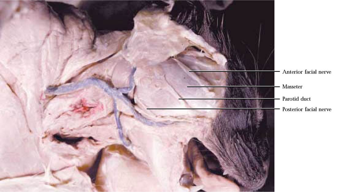
FIGURE 2-21 Jaw muscle I.
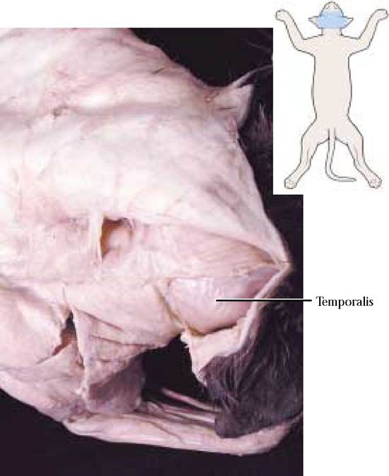
FIGURE 2-22 Jaw muscle II.
SHOULDER MUSCLES
Supraspinatus m.
This thick muscle lies in the supraspinous fossa of the scapula [Figure 2-19].
Origin: From the entire surface of the supraspinous fossa Insertion: Greater tuberosity of the humerus
Action: Protracts the humerus
Infraspinatus m.
This thick, but somewhat smaller muscle fills the infraspinous fossa [Figure 2-19].
Origin: From the surface of the infraspinous fossa
Insertion: Lateral surface of the greater tuberosity of the humerus
Action: Rotates the humerus laterally
Teres major m.
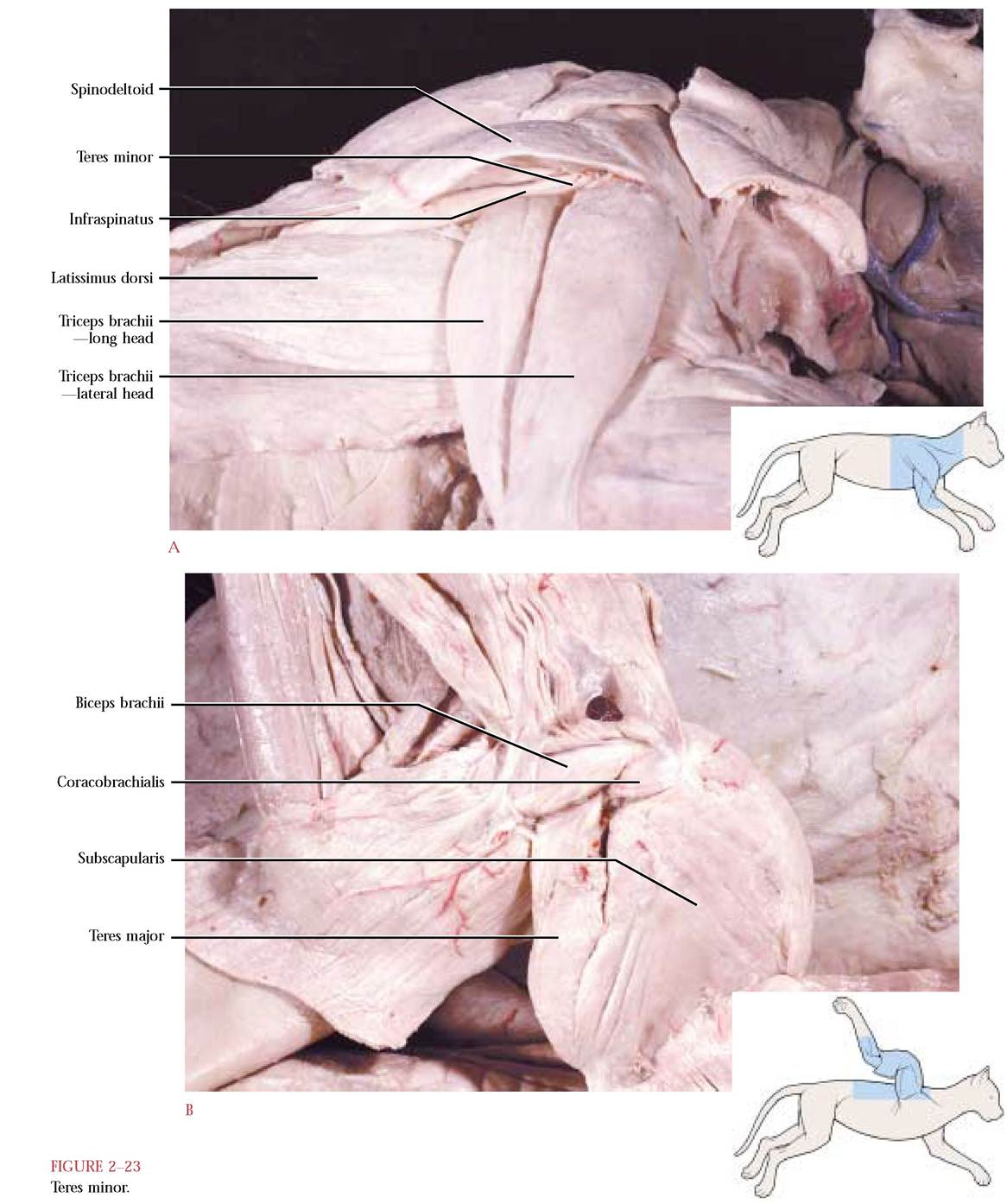
This is a thick, triangular muscle occupying the axillary border of the scapula [Figure 2-19 and Figure 2-23B]. Carefully separate the teres major from the infraspinatus.
Origin: Dorsal third of the axillary border
Insertion: Common tendon with the latissimus dorsi onto the medial surface of the shaft of the proximal end of the humerus
Action: Flexes and rotates the humerus medially
Levator scapulae ventralis m.
This is a band-like muscle whose cranial end emerges from beneath the clavotrapezius and a part of which can be seen lying between the clavotrapezius and acromiotrapezius muscles [Figure 2-13 and Figure 2—14]. This muscle is not present in humans.
Origin: By two heads from the ventral surface of the transverse process of the atlas and from the basioccipital near the tympanic bulla
Insertion: The two heads unite forming a flat band that inserts onto the ventral border of the metacromion of the scapula and into the infraspinous fossa
Action: Pulls scapula craniad
Acromiodeltoid m.
This flat muscle is positioned ventral to the levator scapulae ventralis and caudal to the clavobrachialis [Figure 2—13 and Figure 2-14].
Origin: Acromion of the scapula
Insertion: Surface of the spinodeltoid muscle
Action: Flexes the humerus and rotates it laterally
Spinodeltoid m.
This muscle lies ventral to the acromiotrapezius and levator scapulae ventralis and caudal to the acromiodeltoid [Figure 2-13 and Figure 2-14]. The human deltoid is equivalent to the clavobrachialis (clavodeltoid), acromiodeltoid and spino- deltoid muscles of the cat.
Origin: Spine of the scapula
Insertion: Deltoid ridge of the humerus
Action: Synergistic action with the acromiodeltoid in flexing the humerus and rotating it outward
Teres minor m.
This is a small, somewhat triangular muscle located between the infraspinatus and long head of the triceps brachii and beneath the spinodeltoid [Figure 2-23A]. To find the teres minor, lift the spinodeltoid, exposing the infraspinatus and the closely adherent teres minor. Look for a very subtle connective tissue line and use a probe to separate the two muscles along this line.
Origin: Axillary border of the scapula near the glenoid fossa
Insertion: Greater tuberosity of the humerus
Action: Synergistic action with the infraspinatus in rotating the humerus laterally
Subscapularis m.
This large, medial, triangular muscle is located in the subscapular fossa [Figure 2-20A and Figure 2-23B].
Origin: Almost the entire subscapular fossa
Insertion: Dorsal border of lesser tuberosity of the humerus
Action: Adducts the humerus
MUSCLES OF THE UPPER ARM OR BRACHIUM
Coracobrachialis m.
A very short, bandlike muscle that lies on the medial aspect of the shoulder joint in close proximity to the insertion of the subscapularis and the origin of the biceps brachii [Figure 2-20A and Figure 2-23B].
Origin: Coracoid process of the scapula
Insertion: Proximal end of humerus
Action: Adducts the humerus
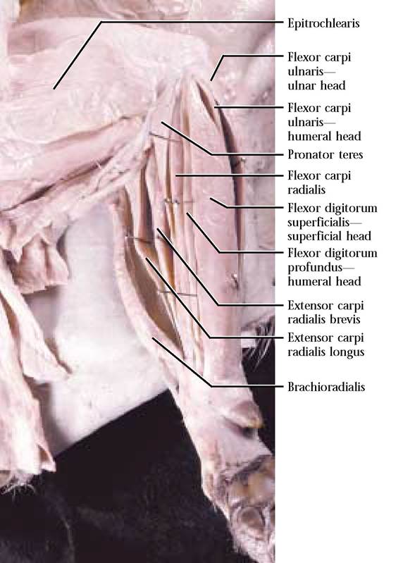
Epitrochlearis m.
This delicate, flat muscle appears on the medial surface of the brachium and partially overlies the triceps brachii [Figure 2-27A]. The muscle has no homolog in man.
Origin: From the lateral border of latissimus dorsi Insertion: By a thin aponeurosis and continuous with the antebrachial fascia onto the olecranon process of the ulna
Action: Acts synergistically with the triceps brachii in extending the antebrachium
Biceps brachii m.
This is a thick muscle lying on the cranial surface of the humerus [Figure 2-20A and Figure 2-23B]. In the human, this muscle has two heads, a long and a short head, while in the cat only the homolog of the long head occurs.
Origin: By a tendon above the glenoid fossa of the scapula
Insertion: By a tendon on the radial tuberosity
Action: Flexes the forearm synergistically with the brachialis, tends to supinate the manus and stabilizes the shoulder joint
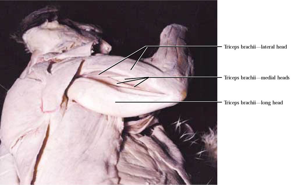
Triceps brachii m.
A very large, lateral muscle consisting of three heads that originate from separate sites, but with a common insertion [Figure 2-24].
Origin: (1) Lateral head—deltoid ridge of proximal end of humerus
(2) Long head—Near glenoid fossa of axillary border of scapula
(3) Medial head—consists of three parts all of which originate from the humerus
Insertion: By a common strong tendon onto the surface of the olecranon process of the ulna
Action: Extends the forearm
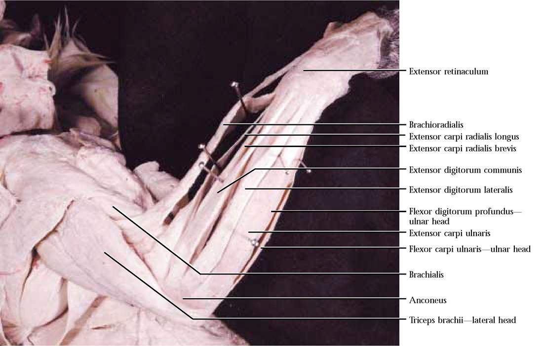
Anconeus m.
This small, triangular muscle covers the lateral surface of the elbow [Figure 2-25].
Origin: Dorsal surface of the lateral epicondyle
Insertion: Lateral surface of the ulna
Action: Acts synergistically with the triceps brachii in extending the forearm
Brachialis m.
A lateral muscle that is located along the cranial surface of the humerus and lies partially obscured by the lateral head of the triceps brachii [Figure 2-25].
Origin: Lateral surface of the shaft of the humerus
Insertion: Lateral surface of ulna near semilunar notch
Action: Flexes the forearm or antebrachium and is synergistic with the biceps brachii.
MUSCLES OF THE FOREARM OR ANTEBRACHIUM
Notice that the antebrachium is covered with a two-layered connective tissue called the antebrachial fascia. The outer layer is loose and is a continuation of the subcutaneous fascia. The inner layer is in close contact with the underlying muscles and extends between the dorsal or extensor muscles and adheres very closely to their tendons. This sheet is continuous on the ventral or flexor surface and is closely attached to the pronator teres and radius. In the carpal area the fascia thickens to form a dorsal transverse ligament, the extensor retinaculum [Figure 2-25] and a ventral transverse ligament, the flexor retinaculum [Figure 2-27B and Figure 27C] to hold the tendons of these muscles in place. It further continues dorsally as the fascia of the manus, while ventrally on the palmar surface it unites with the pad and is continuous with the tendon sheaths of the flexors.
At the level of the first phalanx, narrow, tough annular ligaments that surround the tendons of the flexor muscles are found in these sheets. During the dissection of the forearm muscles it is imperative to trace tendons of each of the muscles to their endpoint. Carefully remove the antebrachial fascia, being especially cautious in the region of the tendons, retinacula and the brachioradialis muscle. This is an area in which one can be destructively creative. Take great care in separating the muscles while maintaining their integrity. In other words, do not split muscles. The brachioradialis, the extensor carpi radialis longus, the extensor carpi radialis brevis and flexor digitorum profundus (ulnar head) muscles can be observed and identified in dorsal and ventral views of the antebrachium. You must be able to recognize them in both views.
For an orderly separation of the extensor muscles, begin the isolation of muscles at the “thumb ” or radial side of the antebrachium.
Brachioradialis m.
This narrow, bandlike muscle extends along the radial border of the antebrachium in company with blood vessels and a nerve [Figure 2-25].
Origin: Mid-shaft of the humerus
Insertion: Styloid process of the radius
Action: Supinates the manus
Extensor carpi radialis longus m.
This is a slender muscle whose main mass lies on the radial side of the antebrachium, deep to the brachioradialis [Figure 2-25].
Origin: Lateral supracondyloid ridge of the humerus
Insertion: Thin tendon at the base of the second metacarpal Action: Extends the manus
Extensor carpi radialis brevis m.
This slender, somewhat shorter muscle, lies just medial to the extensor carpi radialis longus and must be carefully separated from it [Figure 2-25].
Origin: Lateral Supracondyloid ridge of the humerus below the origin of the extensor carpi radialis longus
Insertion: Tendon at the base of the third metacarpal
Action: Extends the manus
Extensor digitorum communis m.
A long, slender, dorsal muscle that partially overlies the extensor carpi longus and brevis [Figure 2—25]. The human muscle is called simply, extensor digitorum.
Origin: Lateral supracondyloid ridge of humerus below the origin of the extensor carpi radialis brevis
Insertion: Tendon divides into four slips that insert on the dorsal surface along the medial aspect of the three phalanges of the second, third, fourth and fifth digits
Action: Extension of second, third, fourth and fifth digits
Extensor digitorum lateralis m.
A long, slender, dorsal muscle that lies lateral to the extensor digitorum communis [Figure 2-25]. Humans lack this muscle, but possess an extensor digiti minimi that inserts on the fifth digit only and extends the little finger.
Origin: Lateral supracondyloid ridge of humerus below the origin of the extensor digitorum communis
Insertion: Division of the tendon similar to that of the extensor digitorum communis, but may subdivide into three or four parts that insert along the dorsolateral surface of the phalanges of digits three, four, and five or two, three, four, and five, respectively
Action: Extends the digits with the extensor digitorum communis, synergistically
Extensor carpi ulnaris m.
This long, slender muscle lies along the ulnar side of the antebrachium and is the last of the superficial extensors [Figure 2-25].
Origin: Lateral epicondyle of the humerus below the origin of the extensor carpi lateralis and ulna above the semilunar notch
Insertion: Base of the fifth metacarpal
Action: Extension of carpals of the ulnar side
To expose the supinator, abductor policis longus and the extensor indicis muscles, reflect the extensor digitorum lateralis medially and the extensor carpi ulnaris laterally.
Supinator m.
This flat muscle surrounds the proximal end of the radius and lies under the extensor digitorum communis and lateralis [Figure 2-26].
Origin: From stabilizing elbow ligaments and the lateral epicondyle of the humerus
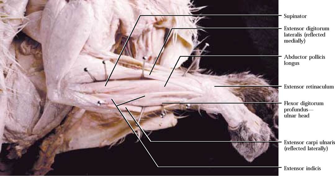
FIGURE 2-26 Deep forearm extensors, supinator, and abductor policis longus.
Insertion: Fibers pass obliquely to insert on the proximal third of the radius
Action: Supinates the forearm synergistically with the brachioradialis
Abductor pollicis longus m.
A flat muscle whose oblique fibers occur between the radius and ulna, is located distal to the supinator [Figure 2-26].
Origin: Ventrolateral surface of the ulnar shaft and the dorsal surface of the radius
Insertion: On the radial side of the first metacarpal
Action: Extends and abducts the pollex (thumb, digit I).
In the human, similar abduction and extension of the thumb involves the abductor pollicis longus and the extensor pollicis brevis, absent in the cat
Flexor carpi radialis m.
A thin muscle that extends from the humerus to the manus
[Figure 2-27A].
Origin: Medial epicondyle of the humerus
Insertion: Bases of the second and third metacarpals
Action: Flexes the wrist
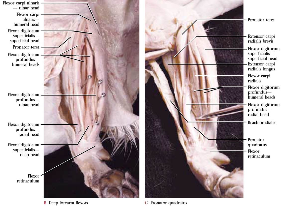
Flexor digitorum profundus m.
This is a deep five-headed muscle whose tendons are united at the wrist [Figure 2-27B and Figure 2-27C].
Origin: Most of the radial border of the ulna (ulnar head), middle third of the radius, interosseous ligament between the radius and ulna and nearby shaft of the ulna (radial head), medial epicondyle of the humerus (three humeral heads)
Extensor indicis m.
This very slender muscle lies deep to the extensor carpi ulnaris. It may consist of a single muscle or two separate muscles with a common origin [Figure 2-26].
Origin: Lateral surface of the ulna
Insertion: At the base of the radius; if the muscle is single with a single tendon, it inserts on the second phalanx of the second digit; if the muscle is single with a divided tendon, both may insert on the second phalanx of the second digit or one may insert on the second digit and one on the pollex (digit I); if there are two muscles, an extensor digiti I, inserts by means of a tendon on the pollex and an extensor digiti II inserts on digit II
Action: Extends the first and second digits; in man, extensor digiti I and extensor digiti II are represented by the extensor pollicis longus and the extensor indicis proprius, respectively
Similar to the extensor muscles, begin the separation of the superficial flexor muscles at the “thumb ” side.
Pronator teres m.
This ventral muscle is oriented obliquely over the upper surface of the forearm [Figure 2-27A and Figure 2-27B]. In the human there are two heads, a humeral head and an ulnar head.
Origin: Medial epicondyle of the humerus
Insertion: Middle of the medial border of the radius
Action: Pronation of the manus by rotating the radius
A Superficial forearm flexors
FIGURE 2-27 Medial forearm muscles.
Insertion: Five tendons join at the wrist to form a strong, wide, white, glistening band that subdivides into five tendons to insert on the bases of the distal phalanx of digits 1-5
Action: Flexes all digits
Flexor digitorum superficialis m.
This is a muscle that occurs in two parts, superficial and deep, that inserts on the digits [Figure 2-27A and Figure 2-27B]. The superficial head is the widest of the flat, bandlike muscles of the ventral surface of the lower forearm. The deep head lies on the surface of the tendon of the flexor digitorum profundus. A palmaris longus m. is present in the human, but not in cats. Some authors, however, describe the superficial portion of flexor digitorum superficialis as the palmaris longus in the cat.
Origin: Superficial head—medial epicondyle of the humerus; Deep head—tendon of two humeral heads of the flexor digitorum profundus
Insertion: The tendons of the two heads pass under the flexor retinaculum and split to insert on either side of the middle phalanx of digits 2-5
Action: Flexes the digits
Flexor carpi ulnaris m.
A muscle having a humeral and ulnar head that lies along the ulnar side of the ventral aspect of the lower forearm [Figure 2-27A and Figure 2-27B].
Origin: Humerus near the medial epicondyle (humeral head), lateral surface of the olecranon of the ulna (ulnar head)
Insertion: Pisiform bone of the carpals
Action: Wrist flexor
MUSCLES OF THE MANUS
Lumbricales m.
Pronator quadratus m.
A deep muscle whose fibers extend obliquely between the distal ends of the ulna and the radius. To find this muscle, separate the tendon and radial head of the flexor digitorum profundus and the tendon of flexor carpi radialis. Look for a flat, bluish purple muscle covered by shiny fascia. Carefully slit the fascia to reveal the oblique fibers of this muscle [Figure 2-27C].
Origin: Distal part of the ventral border of ulna
Insertion: Distal surface of the ventral border of the radius
Action: Rotates the radius and is synergistic with the
pronator teres
These are small intrinsic muscles of the manus.
Origin: By slips from the common tendon of flexor digitorum profundus
Insertion: Base of the first phalanx of digits 2-5
Action: Bends digits radially
A number of additional small intrinsic muscles associated with the metacarpals and digits will not be treated here. You might wish to examine them independently.
MUSCLES OF THE THIGH
Sartorius m.
This is a muscle that appears as a narrow, thick band laterally. This is deceiving, however, because it continues as a
thin band extending almost halfway across the medial surface of the cranial aspect of the thigh [Figure 2-28, Figure 2-29, and Figure 2-30]. Use caution in dissecting this muscle since it often has fat and extensive connective tissue associated with it.
Origin: Crest and ventral border of the ilium
Insertion: Patella, tibia, and fascia of the knee
Action: Adducts and rotates the femur, extends the shank
**Sew and cut this muscle.
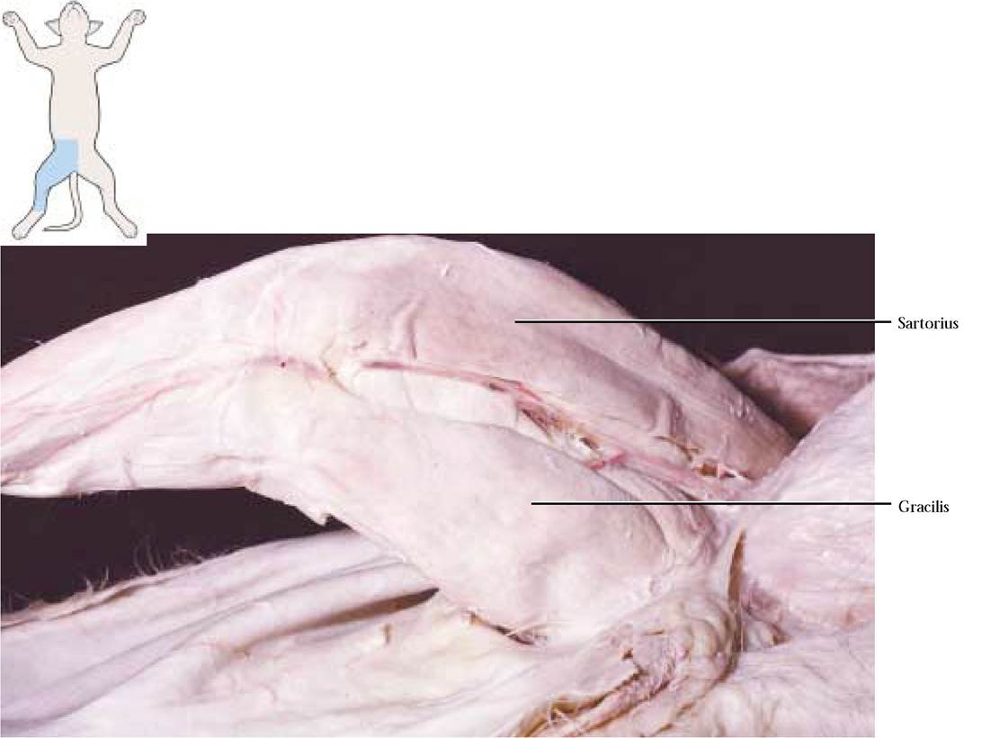
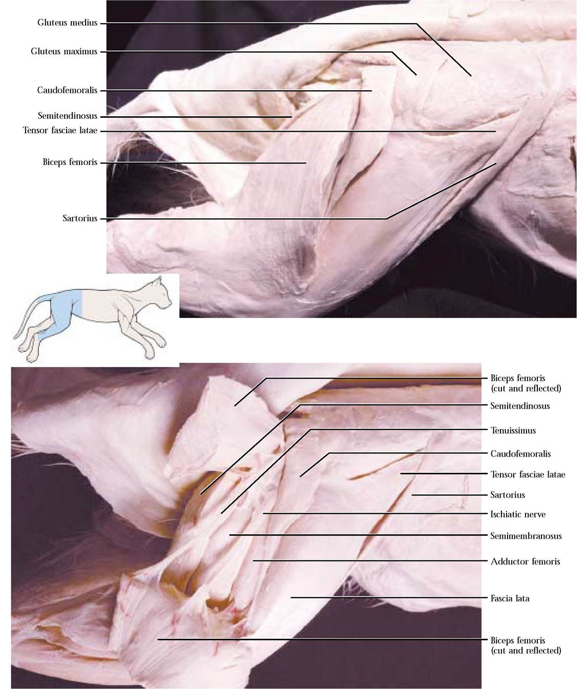
FIGURE 2-29 Lateral thigh muscles.
Gracilis m.
This is another thin muscle that occupies the caudal half of the medial surface of the thigh [Figure 2-28]. Since the insertion is an aponeurosis, you may tend to be overzealous in cleaning the surface of this muscle and destroy it.
Origin: Symphysis of the ischium and pubis
Insertion: A thin aponeurosis on the medial surface of the tibia and continuous with the fascia of the shank
Action: Adducts and retracts the leg
**Sew and cut this muscle.
Biceps femoris m.
This large, thick muscle covers almost three-fourths of the lateral surface of the thigh [Figure 2-29 and Figure 2-30]. Like the gracilis, care must be exercised when dissecting near the insertion. This muscle will only be cut and not sewn. There are three major concerns when bisecting the biceps: (1) the very delicate tendon of the caudofemoralis is closely applied to the undersurface of the cranial aspect of the biceps, (2) the tenuissimus lies just beneath the caudal edge of the biceps, and (3) the ischiatic nerve is positioned under and approximately in the middle of the biceps. Avoid cutting any of these. This is another muscle, similar to the biceps brachii, that was first described in the human where it originates as two heads (bi-ceps), one from the ischial tuberosity and the second from the femur, while in other mammals, e.g., the cat, it possesses a single head.
Origin: Ischial tuberosity
Insertion: Proximal one-third of the tibia and lateral patella Action: Abducts thigh and flexes the shank
** Only this muscle will be cut. Be extremely careful to avoid severing the underlying tenuissimus, sciatic nerve, and tendon of the caudofemoralis.
Tenuissimus m.
This is an extremely slender muscle that is strongly adherent to the biceps femoris [Figure 2-30]. This muscle is absent in humans.
Origin: Transverse process of second caudal vertebra Insertion: In common with the biceps femoris
Action: Synergistically assists the biceps femoris in abducting the thigh and flexing the shank
Caudofemoralis m.
Just cranial to the biceps femoris, with its major mass lying beneath that muscle, is the caudofemoralis [Figure 2-29 and Figure 2-30]. Again, care must be exercised when dissecting this muscle since it is often associated with fat and connective tissue and is inserted by way of a very thin, narrow tendon that adheres closely to the medial surface of the biceps femoris. The caudofemoralis is missing in humans.
Origin: Transverse processes of second and third caudal vertebrae
Insertion: Thin tendon along the lateral border of the patella
Action: Abducts the thigh and extends the shank
The semitendinosus, the semimembranosus, and the adductor femoris muscles can be observed and identified in both lateral and medial views of the thigh. You will be expected to identify them in both views.
Semitendinosus m.
This is a long muscle that forms the caudal border of the thigh [Figure 2-30 and Figure 2-31].
Origin: Ischial tuberosity
Insertion: Medial surface of the tibia
Action: Flexes the shank
Semimembranosus m.
A thick muscle, cranial to the semitendinosus and lying on the medial aspect of the thigh, is the semimembranosus [Figure 2-30 and Figure 2-31].
Origin: Ischial tuberosity and ramus of the ischium
Insertion: Medial epicondyle of the femur and adjacent medial surface of the tibia
Action: Extends the thigh
Adductor femoris m.
This broad muscle lies cranial to and partially covered by the semimembranosus [Figure 2-30 and Figure 2-31].
Origin: Ramus of pubis and ischium
Insertion: Shaft of femur
Action: Adducts thigh
Adductor longus m.
Cranial to the adductor femoris is the thin adductor longus that appears as a triangle that is just caudal to the femoral vessels and saphenous nerve, all of which lie in the ilio- pectineal fossa [Figure 2-31].
Origin: Craniomedial border of the pubis
Insertion: Middle portion of the linea aspera of the femur
Action: Adducts thigh
Pectineus m.
This small, triangular muscle lies just beneath the femoral vessels and saphenous nerve [Figure 2-31]. Often, fat may obscure the pectineus and will have to be carefully removed.
Origin: Cranial border of pubis
Insertion: Proximal shaft of the femur
Action: Adducts the thigh
ILIOPSOAS COMPLEX
This complex consists of a thick muscle mass that is almost entirely obscured by the abdominal musculature and includes the iliacus and psoas major. To observe, make a two inch lateral incision through the abdominal musculature.
Iliacus m.
The lateral and slightly dorsal portion of the complex is the iliacus [Figure 2-31].
Origin: Ventral border of the ilium
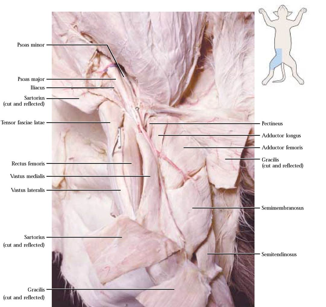
FIGURE 2-31 Thigh muscles, muscles of the iliopsoas complex, psoas minor.
Insertion: Lesser trochanter of the femur
Action: Flexes and rotates the thigh
Psoas major m.
This is the largest and medial portion of this complex [Figure 2-31].
Origin: Bodies of the last thoracic and all of the lumbar vertebrae
Insertion: Lesser trochanter of the femur
Action: Flexes and rotates the thigh
Psoas minor m.
This is a very thin muscle occurring medial to the psoas major [Figure 2-31]. This muscle is missing in a high percentage of humans.
Origin: Bodies of the last thoracic and first few lumbar vertebrae
Insertion: Pubis, by a long, narrow, conspicuous glistening tendon
Action: Flexes vertebral column
Quadratus lumborum m.
This is a deep, flat muscle that lies ventral to the lumbar portion of the vertebral column. This muscle may be best observed during the dissection of the viscera.
Origin: From the last rib and last few thoracic and lumbar vertebrae
Insertion: On the transverse processes of the lumbar vertebrae and on part of the ilium
Action: Bends the vertebral column laterally
Tensor fasciae latae m.
This rather thick, triangular muscle largely covers the cranial portion of the vastus lateralis and abuts the caudal border of the sartorius and the cranioventral border of the gluteus medius [Figure 2-29, Figure 2-30, and Figure 2-31]. Extreme care should be exercised in loosening the fascia lata from the caudal edge of the vastus lateralis. Do not shred or destroy the fascia lata.
Origin: Ventral border of the ilium, fascia of surrounding hip muscles
Insertion: Into the fascia lata. The fascia lata continues distally and covers part of the vastus lateralis and vastus medialis and then inserts on the surface of the patella
Action: Tightens the fascia lata and helps to extend the shank
QUADRICEPS COMPLEX
Four component muscles make up this complex that lies on the craniolateral and craniomedial aspects of the thigh and is the powerful extensor of the shank.
Vastus medialis m.
This is the most medial of the four muscles [Figure 2-31 and Figure 2-32].
Origin: Shaft of the femur
Insertion: Crosses the patella and inserts by means of the patellar ligament on the tibial tuberosity
Action: Extends the shank
Vastus lateralis m.
This is a large, flat muscle that covers the cranial and lateral surface of the thigh [Figure 2-31 and Figure 2-32].
Origin: Shaft and greater trochanter of the femur
Insertion: In common with the vastus medialis and rectus femoris
Action: Extends the shank
Rectus femoris m.
This is a spindle shaped muscle that rests between the vastus medialis and the vastus lateralis and is distinguished by a shiny covering of fascia [Figure 2-31 and Figure 2-32].
Origin: From the ilium near the acetabulum
Insertion: In common with the vastus medialis and lateralis
Action: Extends the shank
Vastus intermedius m.
This is the deepest of the quadriceps muscles and can be seen by carefully separating the rectus femoris and vastus lateralis [Figure 2-32].
Origin: Almost the entire shaft of the femur
Insertion: In common with the other three members of this complex
Action: Extends the shaft
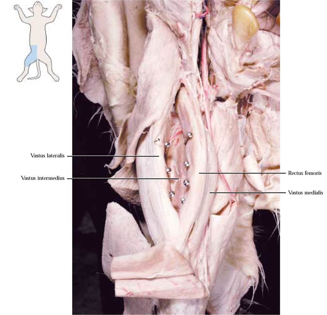
FIGURE 2-32 Quadriceps femoris complex.
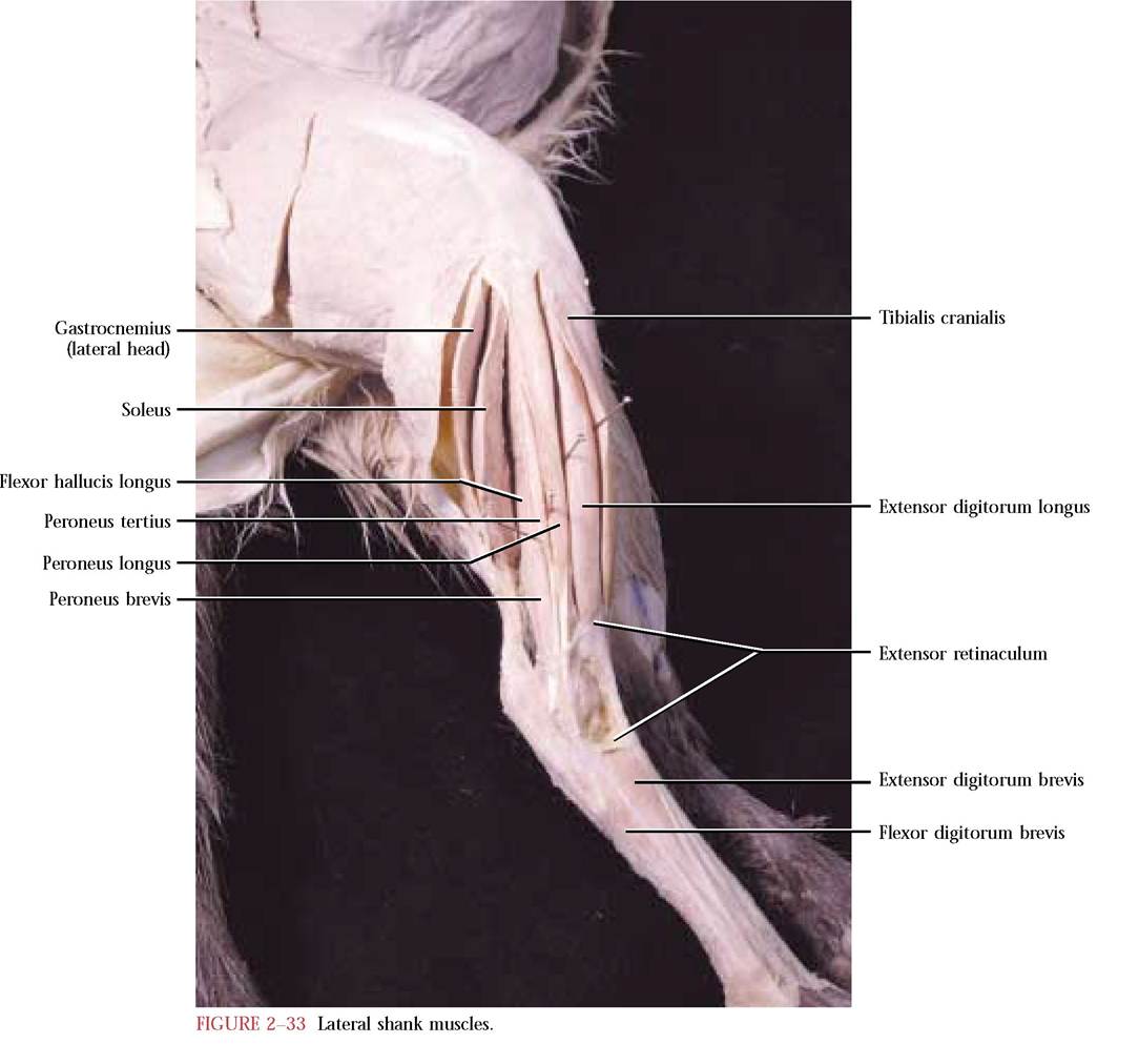
MUSCLES OF THE SHANK
Beware of the tough fascia surrounding the shank muscles. Carefully remove this tissue and while doing so, avoid the destruction of the tendons of insertion of the thigh muscles. The shank muscles are roughly divided into extensors and flexors. A distinct band of connective tissue, the extensor retinaculum, holds the cranial extensor muscle tendons in place [Figure 2-33].
In the following actions of the shank muscles, extension of the pes is synonymous with plantar flexion and flexion of the pes is synonymous with dorsiflexion. Flexion of a shank muscle decreases the angle of the joint at the ankle by pulling the pes closer to the shank and extension of a shank muscle increases the angle of the joint at the ankle by pulling the pes away from the shank.
Tibialis cranialis m.
This large, flat, meaty muscle lies on the craniolateral aspect of the tibia [Figure 2-33].
Origin: Proximal end of the tibia and fibula
Insertion: Along the medial surface of the first metatarsal after passing beneath the extensor retinaculum
Action: Flexes the pes
Extensor digitorum longus m.
This large muscle lies beneath the tibialis cranialis and only a narrow strip is evident caudal to it on the lateral surface of the shank [Figure 2-33]. Carefully loosen this muscle from the tibialis cranialis.
Origin: Lateral epicondyle of the femur
Insertion: The tendon, after passing under the extensor retinaculum, is subdivided into four slips that insert on the dorsal surface of the second and third phalanges of digits 2-5
Action: Extends the digits and flexes the pes
Extensor hallucis longus m.
This is a muscle lying deep to the tibialis cranialis and the extensor digitorum longus.
Origin: Anterior surface of the fibula
Insertion: Generally in common with the tendon of the tibialis cranialis on the first metatarsal
Action: Flexes the pes
Peroneus longus m.
This is one of three peroneus muscles on the lateral surface of the shank. It is the most superficial of this group [Figure 2-33].
Origin: Head and lateral surface of the shaft of the fibula
Insertion: Proximal ends of all five metatarsals
Action: Extends the pes
Peroneus tertius m.
This is a slender muscle lying beneath the peroneus longus [Figure 2-33].
Origin: Lateral surface of the fibula
Insertion: Its tendon lies in the groove of the lateral malleolus and passes along the lateral margin of the foot and inserts on the first phalanx of the fifth digit.
Action: Flexes pes and abducts and extends fifth digit
Peroneus brevis m.
The shortest of the three peroneus muscles, peroneus brevis, lies posterior to the other two [Figure 2-33].
Origin: Distal half of the fibula
Insertion: *Note that the tendon of peroneus brevis passes in common with the tendon of peroneus tertius within the groove of the lateral malleolus and finally inserts on the base of the fifth metatarsal on the lateral side
Action: Extends the pes
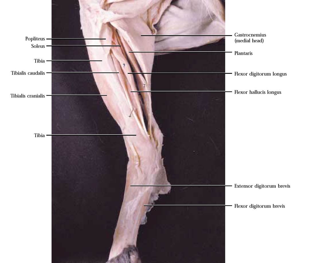
Popliteus m.
This is a triangular muscle that wraps obliquely around the posterior aspect of the knee from the femur to the tibia [Figure 2-34]. It appears as a flat, triangular wedge of muscle overlying the origins of the shank flexor muscles, all of which are obscured by the tendon of insertion of the semitendinosus. To observe the popliteus, carefully separate the tendon of semitendinosus and lift it toward the thigh. The fibers of popliteus lie obliquely over the perpendicular fibers of the shank flexors. Use a probe to free the surface of the edge of popliteus from the underlying flexors.
Origin: Lateral epicondyle of the femur
Insertion: Medial aspect of the proximal end of the tibia
Action: Flexes and medially rotates the leg
Flexor digitorum longus m.
This is a long, slender muscle on the medial aspect of the shank just posterior to the tibia [Figure 2-34].
Origin: Head of the fibula and shaft of the tibia
Insertion: Forms a common broad tendon extending over the plantar surface of the foot, then dividing into
four discrete tendons that insert on the base of the terminal phalanx of each toe
Action: Flexes toes and pes
Flexor hallucis longus m.
This somewhat larger muscle lies lateral to the flexor digitorum longus on the posterior aspect of the shank. [Figure 2-33 and Figure 2-34].
Origin: From the shaft of the fibula and shaft of the tibia
Insertion: In common with the flexor digitorum longus
Action: Flexes toes and pes
Tibialis caudalis m.
This is a flat, slender muscle that lies beneath the flexor digitorum longus and between the flexor digitorum longus and flexor hallucis longus [Figure 2-34]. Notice the prominent tendon of this muscle.
Origin: From the head of the fibula, proximal end of the tibia and adjacent aponeurosis
Insertion: By means of a long slender tendon onto the plantar surface of the navicular and medial cuneiform
Action: Extensor of the pes
Gastrocnemius m.
The gastrocnemius is the major contributor to most of the bulky posterior muscle mass of the lower leg known as the calf [Figure 2-33 and Figure 2-34]. It possesses two heads, a lateral and a medial head.
Origin: The lateral head arises from the lateral border of the patella, the superficial fascia of the shank, the sesamoid bone located above the lateral epicondyle of the femur and an aponeurosis from the plantaris and adjacent tibia. The medial head originates from the sesamoid bone above the medial epicondyle of the femur and its distal adjacent shaft.
Insertion: By means of a common powerful tendon, the Achilles tendon, formed by the individual tendons of the gastrocnemius, the soleus, and the plantaris muscles, that inserts on the proximal end of the calcaneus
Action: Extends the pes
Plantaris m.
The plantaris lies beneath the gastrocnemius and can be seen protruding between the proximal ends of the heads of this muscle [Figure 2-34]. Care must be exerted in dissecting this muscle away from the two heads of the gastrocnemius.
Origin: From the sesamoid above the lateral epicondyle of the femur and the lateral border of the patella Insertion: Passes through the center of the Achilles tendon over the calcaneus and serves as the origin of the flexor digitorum brevis on the ventral aspect of the pes
Action: Acts synergistically with the gastrocnemius and the soleus to extend the pes
Soleus m.
This is a flat muscle located beneath the plantaris [Figure 2-33 and Figure 2-34].
Origin: Proximal part of the fibula
Insertion: In common with the tendon of the gastrocnemius and contributes to the formation of the Achilles tendon
Action: Synergistic extension of the pes with the gastrocnemius and plantaris
Triceps surae m.
The gastrocnemius and the soleus muscles have sometimes been considered a calf muscle with three heads, known as the triceps surae.
MUSCLES OF THE HIP
Carefully remove the tough fascia covering the hip region. Reidentify the tensor fasciae latae, the caudofemoralis and the biceps femoris [Figure 2-29]. Do not destroy nerves in this area.
Gluteus maximus m.
The gluteus maximus is a thin trapezoidal muscle lying just anterior to the caudofemoralis [Figure 2-29]. In humans, this muscle is massive and makes the major contribution to nice “buns. ”
Origin: From transverse processes of the last sacral and first caudal vertebrae, as well as adjacent fascia
Insertion: Onto the surface of the greater trochanter of the femur
Action: Abducts the thigh
**After this muscle has been identified and isolated, slide a probe under the muscle perpendicular to the fibers in the region of the belly and with a sharp scalpel cut along the top of the probe, using the probe as a hard surface to prevent cutting into important muscles beneath it. Do not sew this muscle.
MUSCLES OF THE PES
Extensor digitorum brevis m.
This thin muscle covers the dorso-lateral surface of the tarsus and metatarsus [Figure 2-33 and Figure 2-34].
Origin: From the proximal ends of metatarsals 3-5 Insertion: By three tendons that split into a medial and lateral slip that both terminate on the dorsal and lateral surface of the first phalanx
Action: Extends the toes
Flexor digitorum brevis m.
This muscle lies on the plantar surface of the foot [Figure 2-33 and Figure 2-34].
Origin: An extension of the plantaris tendon
Insertion: By four tendons to the second phalanx of digits 2-5
Action: Flexes the toes
In addition, there are a number of small muscles associated with the tarsus, metatarsus and phalanges that will not be individually described. Various intricate movements are caused by them.
Gluteus medius m.
This muscle, in the cat, is a very thick muscle sandwiched between the cranial tensor fasciae latae and caudal gluteus maximus [Figure 2-29].
Origin: From the crest and lateral surface of the ilium, transverse processes of the last sacral and first caudal vertebrae and adjacent fascia
Insertion: Proximal end of the greater trochanter of the femur
Action: Abducts the thigh
** Extreme care must be exercised in dissecting this muscle from the important, underlying musculature with which you will be dealing, shortly. After cutting and reflecting the gluteus maximus muscle, you should be able to identify the flattened posterior portion of the pyriformis extending from beneath the caudal edge of the gluteus medius muscle. Note the ischiatic nerve passing obliquely under the pyriformis muscle. The cranial edge of the gluteus medius is very thick and must be loosened from the muscular part of tensor fasciae latae. Lift the cranial edge of the gluteus medius and observe the shiny, spindle-shaped gluteus minimus muscle. Insert a probe beneath the caudal edge of the gluteus medius and perpendicular to the direction of the muscle fibers, continuing until it emerges beneath the cranial edge. Be careful that the probe remains dorsal to the pyriformis and gluteus minimus muscles. With a sharp scalpel, cut through the belly of the gluteus medius along the dorsal edge of the probe. Do not sew this muscle.
Pyriformis m.
This fan-shaped muscle lies under both of the superficial gluteus muscles [Figure 2-35A and Figure 2-35B]. As mentioned earlier, notice that the ischiatic nerve passes under this muscle.
Origin: From transverse processes of the last two sacral and first caudal vertebrae
Insertion: Proximal end of the greater trochanter of the femur
Action: Abducts the thigh
**Insert a probe perpendicular to the fibers of the pyriformis approximately 3/4 of the distance from the point of origin, beneath the caudal edge until it emerges cranially. Take care not to pick up the ischi- atic nerve with the muscle. With a sharp scalpel, cut through the pyriformis, following the top edge of the probe. Do not sew this muscle.
Gluteus minimus m.
This muscle consists of an anterior shiny, spindle-shaped portion and a flat, fan-shaped posterior portion [Figure 2-35A and Figure 2-35B].
Origin: From the lateral surface of the ilium
Insertion: Greater trochanter of the femur
Action: Abducts and rotates the thigh outward
Articularis coxae m.
This is a small, flat muscle lying beneath gluteus minimus and between the heads of the vastus lateralis and rectus femoris [Figure 2-35C]. It does not exist in humans.
Origin: Ilium cranial to the acetabulum
Insertion: Dorsal surface at the proximal end of the femur Action: Assists in flexion and rotation of the thigh
Gemellus cranialis m.
This is a fan-shaped muscle lying below the pyriformis and closely abutting the caudal border of the gluteus minimus [Figure 2-35B].
Origin: From the dorsal edge of the ilium and ischium
Insertion: Area anterior to the apex of the greater trochanter of the femur
Action: Abducts and rotates the thigh
Coccygeus m.
This is a deep hip muscle lying dorsal to gemellus cranialis and often mistaken as part of it [Figure 2-35B].
Origin: In common with gemellus cranialis, the dorsal edge of the ilium and ischium
Insertion: Proximal caudal vertebrae
Action: Contributes to the pelvic wall and bends the tail laterally.
Obturator internus m.
This muscle is located directly caudal to the gemellus cranialis [Figure 2-35B]. Although in lateral view, it appears as a small triangular muscle, its actual area is considerably more extensive.
Origin: Along the medial surface of the ramus of the ischium from the symphysis to the tuberosity
Insertion: The muscle passes laterally over the dorsal border of the ischium to insert into the trochanteric fossa of the femur
Action: Abducts the thigh
Gemellus caudalis m.
This flat muscle lies caudal to and is overlapped by the obturator internus [Figure 2-35B].
Origin: From the dorsolateral surface of the ischium
Insertion: In common with the tendon of the obturator internus into the trochanteric fossa
Action: Abducts the thigh
Quadratus femoris m.
The last of the visible deep hip muscles is short and thick and frequently appears brownish [Figure 2-35B].
Origin: Lateral surface of the ischial tuberosity
Insertion: Along the ventral borders of the greater and lesser trochanters
Action: Retraction of the thigh
Obturator externus m.
Even though one might expect this muscle to be easily visible from the external surface, it actually is deeply buried beneath
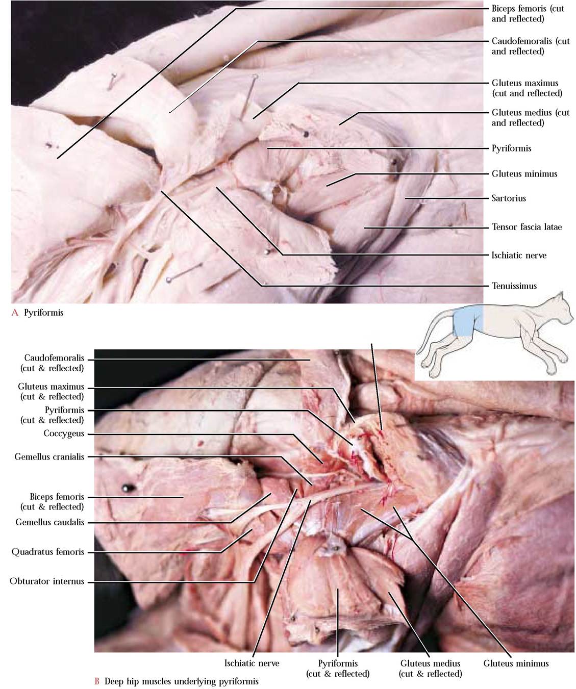
Gluteus medius (cut & reflected)
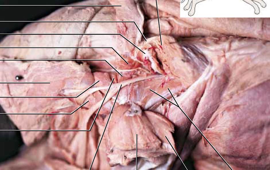
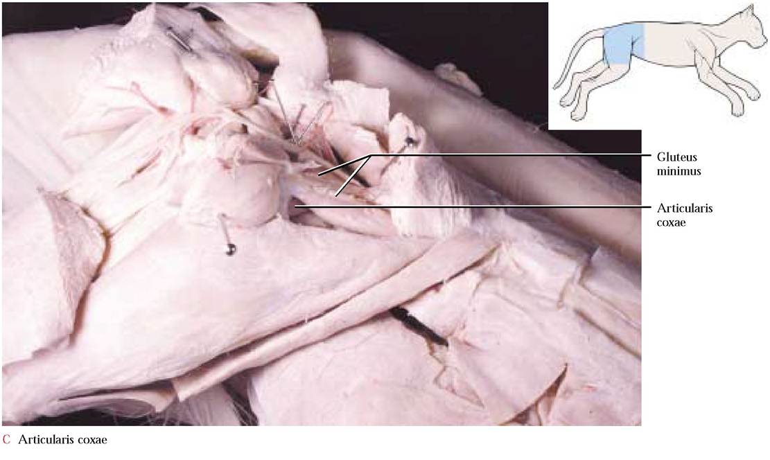
some of the hip muscles already studied. To observe this muscle, carefully separate the gemellus caudalis and quadratus femoris muscles.
Origin: From the lateral surface of the pubis and ischium near the border of the obturator foramen
Insertion: Into the trochanteric fossa of the femur
Action: Rotates and retracts the thigh
TAIL MUSCLES
A number of intrinsic muscles perform the typical tail movements of a feline, e.g., straight carriage, lateral bending and the nervous twitching and lashing motions prior to pouncing upon some unsuspecting object. Several of these are continuations of the lumbar muscles discussed previously, e.g., the extensor caudae medialis, a continuation of the multifidus spinae, raises the tail.