» Nervous System
The brain and spinal cord do not differ appreciably from the usual pattern. The difference in the spinal accessory nerve has been previously noted. Few other studies on specific cranial nerves of the llama or alpaca are available, and function of the cranial nerves can be assessed in the traditional manner.
As in other species, the facial nerve is relatively superficial and can be damaged from trauma peripherally, or from inflammation in its passage through the skull.The spinal cord ends at the second sacral vertebra. The basic distribution of the peripheral nerves is presumed to be similar to other species as well, although specific anatomic studies on the peripheral nerves of the llama and alpaca are lacking, and evidence suggests that the nerves to the distal limb differ somewhat from the symmetrical medial/lateral pattern of the horse and ruminant. In contrast to the situation in horses and ruminants, the major nerve supply to the foot of the South American camelid is on only the medial aspect of the metatarsus or metacarpus.
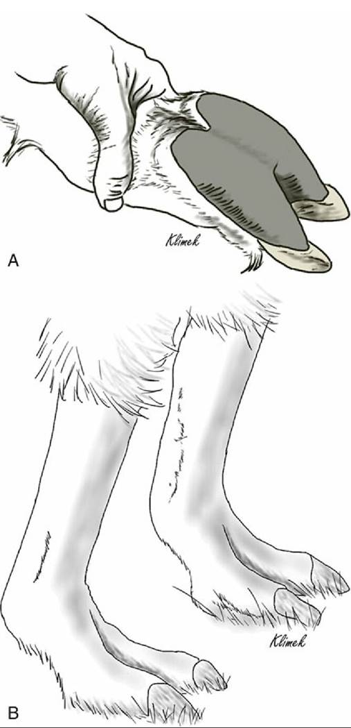
FIG. 38.39 Palmar (A) and dorsal (B) views of the front foot of a llama.
Epidural Anesthesia and Collection of Cerebrospinal Fluid: Regional nerve blocks for the limbs are used less in llamas and alpacas than in other species because the path of individual nerves is not well described. Epidural anesthesia can be performed in camelids. The caudal location for perineal procedures is determined by moving the tail and palpating for the first movable joint. In most individuals, the five sacral vertebrae are fused, and the first freely movable joint is S5-C1. However, in some animals, S5 is free but the joint space between S4 and S5 is small enough that injection is usually difficult. If difficulty is encountered, one should move to the next most caudal movable joint.
Because camelids have little negative pressure in the epidural space, the method of inserting only the needle and watching for a drop of anesthetic to be pulled in, is unreliable in these species, and instead the syringe should be left attached to the needle and anesthetic injected when resistance to pressure on the plunger is detected. One must be aware of the fact that the anesthetic may travel cranially enough to involve the lumbosacral nerve roots and cause recumbency. Lumbosacral epidural anesthesia is also possible for abdominal and orthopedic procedures; one must again avoid cranial spread of the anesthetic agent to the level of the brachial plexus and phrenic nerve.Collection of cerebrospinal fluid is similar to procedures used for cattle and horses, and either the atlantooccipital space or the lumbosacral space may be used.
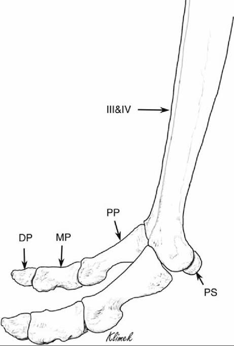
FIG. 38.40 Hindfoot of a camel. DP, Distal phalanx; MP, middle phalanx; PP, proximal phalanx; PS, proximal sesamoid. III&IV, Fused third and fourth metatarsals.