Organization of the Gonads Is Under Genetic Control
The initial development of the embryonic ovary involves the migration of germ cells into the genital ridge from the yolk sac. These primordial germ cells populate sex cords that have formed in the cortical region of the embryonic gonad from the proliferation of cells from the coelomic epithelium (so- called germinal epithelium) of the genital ridge.
The sex cords contribute cells, known initially as follicle cells and subsequently as granulosa cells, which immediately surround the oocyte. The mesenchyme of the genital ridge contributes cells that will become the theca. The entire structure is called a follicle, which includes oocyte, granulosa, and theca cells.No direct connections are formed between the oocytes and the tubes destined to become the oviducts, which are derived from Hiiillerian ducts. The final result is that oocytes are released through the surface of the ovary by rupture of tissue elements that surround the ovary; this process is called ovulation. A specialized end of the oviduct, the fimbria, develops to enable the oocyte to be removed efficiently from the surface of the ovary. In some animals, oocytes are tunneled to the fimbria through the use of a bursa, which tends to encompass the ovary; oocytes are directed to a relatively small opening in the bursa.
The development of the embryonic testis is similar to that of the ovary: germ cells migrate into the genital ridge and populate sex cords that have formed from an invagination of the surface (coelomic) epithelium (Figure 35-1). Sertoli cells (male counterparts of granulosa cells) develop from the sex cords, and Leydig cells (male counterparts of thecal cells) develop from the mesenchyme of the genital ridge. One
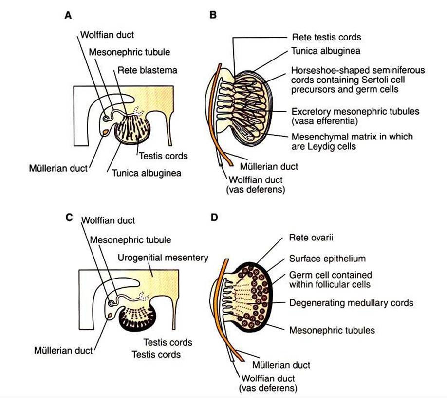
FIGURE 35-1 Testicular development during the 8th week (A) and the 16th to 20th weeks (B) of human fetal life. A,The primitive sex cords proliferate in the medulla and establish contact with the rete testis.The tunica albuginea (fibrous connective tissue) separates the testis cords from the coelomic epithelium and eventually forms the capsule of the testis.
B, Note the horseshoe shape of the seminiferous cords and their continuity with the rete testis cords. The vasa efferentia, derived from the excretory mesonephric tubules, connect the seminiferous cords with the wolffian duct (see text). Comparable diagrams of ovarian development around the 7th week (C) and the 20th to 24th weeks (D) of development. C, Any primitive medullary sex cords degenerate and are replaced by the well-vascularized ovarian stroma.The cortex proliferates, and mesenchymal condensations later develop around the arriving primordial germ cells. D, In the absence of medullary cords and a true persistent rete ovarii, no communication is established with the mesonephric tubules.Therefore, in the adult, ova are shed from the surface of the ovary and are not transported by tubules to the oviduct. (From Johnson M, Everitt B, editors: Essential reproduction, ed 3, London, 1988, Blackwell Scientific.)SECTION Vl Reproduction and Lactation
fundamental difference from ovarian development is that the invagination of the sex cords in the male continues into the medulla of the embryonic gonad, where connections are made with medullary cords from the mesonephros (primitive kidney). The duct of the mesonephros (wolffian duct) becomes the epididymis, vas deferens, and urethra, which has a direct connection to the seminiferous tubules. Thus, male germ cells pass to the exterior of the animal through a closed tubular system.