Outer and Middle Ears Funnel Sound Waves to the Cochlea
The outer ear, composed of the fleshy part (pinna) and the ear canal, funnels sound waves to the tympanic membrane, or eardrum (Figure 17-2). Some animals can move the pinna to more effectively collect sound waves, and the natural shape of the pinna can act to filter selectively certain sound frequencies.
The eardrum is a membrane between the outer and the middle ear. The middle ear is an air-filled cavity in the temporal bone and is connected to the nasopharynx by the auditory (eustachian) tube. Three tiny bones—the malleus, incus, and stapes—collectively called the ossicles, are connected to each other and are located in the middle ear. The malleus is connected to the eardrum, and the stapes is connected to the oval window, a membranous separation between the middle and the inner ear. The ossicles transfer vibration of the eardrum to the oval window in a manner that avoids a significant loss of energy as the sound wave is transferred from the air-filled outer ear to the fluid-filled inner ear. Two small skeletal muscles are also located in the middle ear, with one attached to the malleus and one attached to the stapes. Their contraction reduces the transfer of vibration between the eardrum and the oval window. This can function to protect the inner ear from very loud sounds.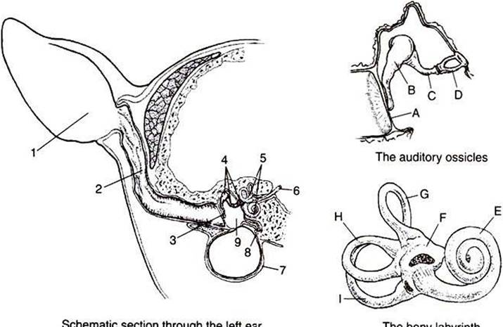
FIGURE 17-2 Schematic sections through the left ear, the auditory ossicles, and the bony labyrinth. 1t Pinna; 2t ear canal; 3t tympanic membrane; 4, auditory ossicles; 5f bony labyrinth; 6t eighth cranial nerve; Ztympanic bulla; 8, eustachian tube; 9t middle ear; At tympanic membrane; Bt malleus; Ct incus; Dt stapes; Et cochlea; Ft utricle; Gt Ht and It semicircular canals. (From Getty R: Atlas for applied veterinary anatomy, cd 2, Ames, 1964, Iowa State University Press.)
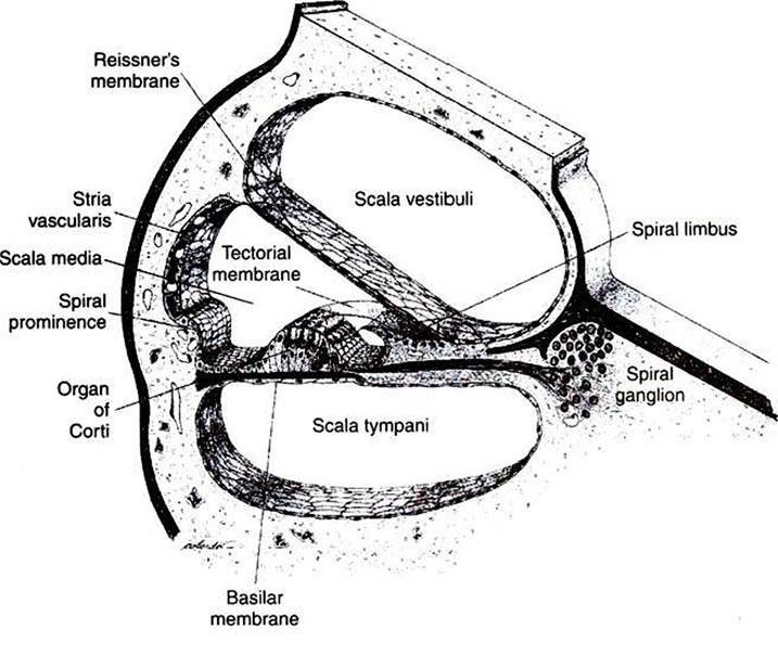
FIGURE 17-3 Schematic representation of a section through one of the turns of the cochlea. (From Bloom W, Fawcett DW: A textbook of histology, ed 10, Philadelphia, 1975, Saunders.)