Parturition and the Puerperal Period: The Neonate
Parturition is initiated mainly by the fetus, although the mother is not without all influence. Mares, for example, tend to give birth when conditions in the stable are quiet and settled.
The endocrine control of birth is complicated and beyond the scope of this book, but certain preparatory changes in the tissues may be mentioned. These take some time to develop, affect many structures, and largely comprise an increase in their water content and loosening of the larger collagen accumulations. The most familiar effect is the in-sinking of the tailhead of cows to the side as parturition impends. Similar but concealed changes soften the caudal reproductive tract, including most significantly the cervix. In some species there is considerable weakening of the pelvic symphysis, but articular changes in domestic animals are limited to some loosening of the sacroiliac joints. After parturition the reproductive organs tend to return toward their former condition, although the restoration after the first pregnancy is never complete. The uterine muscle contracts directly after delivery, and this organ loses much of the weight it gained during pregnancy within a few days.Before this chapter is concluded, a few sentences may be devoted to the status of the newborn, which exhibits interspecific differences that are both striking and important. Neonates of so-called precocial species possess a remarkable ability to fend for themselves more or less at once (Fig. 5.72), whereas those of altricial species are initially much more reliant on maternal care and the warmth and protection of a nest (Fig. 5.73). The young of the ungulate orders, both perissodactyls and artiodactyls, are generally precocial; those of carnivores and primates, including human infants, are predominantly less developed. Young rodents are divided between the two categories; those like rats (myomorphs) are born naked, unable to maintain body temperature independently, and barely capable of struggling to reach the dam's teats, and their eyelids are joined and external ear canals closed by epithelial fusion.
In contrast, guinea pigs and their close relatives (caviomorphs) are born fully haired, mobile, equipped with vision and hearing, and able to seek and ingest solid food within hours of being born (although they may take milk during the first 2 or 3 weeks). The differences among domestic species are significant if less extreme. Foals, like most newborn ungulates, are able to stand and attend their mothers almost at once; their skeletons are well developed, and most secondary ossification centers are not only present but also well advanced in modeling toward their adult form. Relatively efficient locomotor coordination allows them to follow the herd or flock within a short time. Kittens and puppies, on the other hand, have skeletons that are less mature, and many ossification centers have yet to make their appearance (Fig. 5.74); the forelimb musculature is sufficiently developed and controlled to enable them to scramble toward the teats, but that of the hindlimbs is less competent and contributes little to this progress. The development of the sense organs is somewhat retarded, and the eyelids do not part until the tenth day or shortly thereafter. These differences in neonatal status are gradually "ironed out" and most mammals—ourselves excluded—show comparable maturity by the end of the usual lactation period.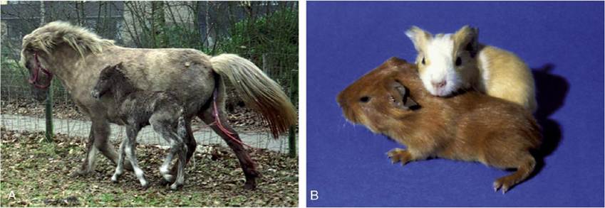
FIG. 5.72 Developmental status shortly after birth. (A) Neonatal foal with mother (the mare has yet to discharge the fetal membranes [afterbirth]). (B) Newborn guinea pigs, which are born in a more developed state.
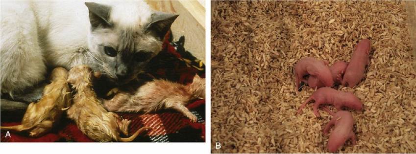
FIG. 5.73 Developmental status shortly after birth in altricial species. (A) Newborn kittens. (B) Three- day-old mouse pups.
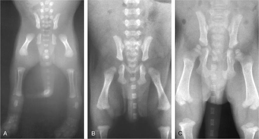
FIG.
5.74 Progress of skeletal ossification in the puppy (A) 1, (B) 14, and (C) 28 days after birth.Comprehension Check
Demonstrate how embryologic development leads to the gross anatomic arrangement of the male reproductive system and facilitate normal sperm development.
Using a dog or a cat cadaver, understand the surgical approach to remove the ovaries and the uterus.
I =I
* Many derivative terms are based on the alternative name, orchis, derived from the Greek.
* The mesorchium is the visceral tunic between the fold (Fig. 5.41) and the epididymal border of the testis but also includes the long peritoneal fold that conveys the testicular vessels and nerves from their origin at the abdominal roof to the testis; it thus forms a considerable portion of the spermatic cord. The narrow fold that attaches the deferent duct to the pelvic and abdominal walls and (more distally) to the mesorchium is the mesoductus deferens.
* Unfortunately, there is some conflict in the use of these terms: many authors reserve uniparous and multiparous for the senses in which we employ monotocous and polytocous.
* The obsolete terms fallopian tubes and oviducts are still encountered, perhaps most commonly in medical writing. Another term, salpinx, receives official recognition; though less frequently encountered, it is the stem of such derivatives as mesosalpinx and salpingitis (inflammation of the uterine tube).
* Compound terms are generally derived from the alternative name, metrium: for example, mesometrium and metritis; surgical removal of the uterus, however, is termed hysterectomy (Greek, hystera, uterus).
* Because many processes continue uninterrupted from one period to the next, there is unavoidable overlap and inconsistency in the use of the terms embryo and fetus.
* In the dog a permanent zone of leaking blood creates marginal hematomas (Fig. 5.67A). In the cat this zone is diffuse and temporary and therefore not as striking as that of the dog.