» Pelvic Limb
The pelvic bones have few remarkable features. Fig. 38.32 shows the skeleton of the pelvic limb of a llama. The obturator foramen is very large and extends laterally to near the acetabulum.
The femur, tibia, and metatarsal bones are all long and slender. The femur lacks a third trochanter. The medial and lateral trochlear ridges of the femur are nearly the same size. The stifle is freer of the trunk than in other species. The stifle is described as having three synovial joint compartments that all communicate, or as having one single compartment, depending on the author. There is one patellar ligament; however, the ligament is broad. The menisci and cruciate ligaments are typical. The lateral collateral ligament has been reported to be absent, with the tendons of origin of the long digital extensor and peroneus tertius acting as support for the joint. The patella is held in place in the trochlear groove by typical femoropatellar ligaments.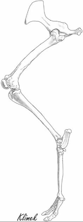
FIG. 38.32 Llama rear limb.
Damage to the femoropatellar ligaments can allow medial or lateral patellar luxation, and upward fixation of the patella can also occur, with the patella catching on the proximal portion of either the medial or the lateral trochlear ridge. Straight-legged ("post-legged") conformation may also cause the patella to ride more proximally, predisposing to upward fixation.
The fibula is reduced to a small projection distal to the lateral condyle of the tibia that is not present in all individuals. The tarsus consists of the talus and calcaneus in the proximal row, the central tarsal bone in the middle row, and the first, fused second and third, and fourth tarsal bones in the distal row. The lateral malleolus of the tibia is a separate bone as in ruminants. The tarsal bones are illustrated in Fig.
38.33. Radiographs of the tarsus are shown in Fig. 38.34.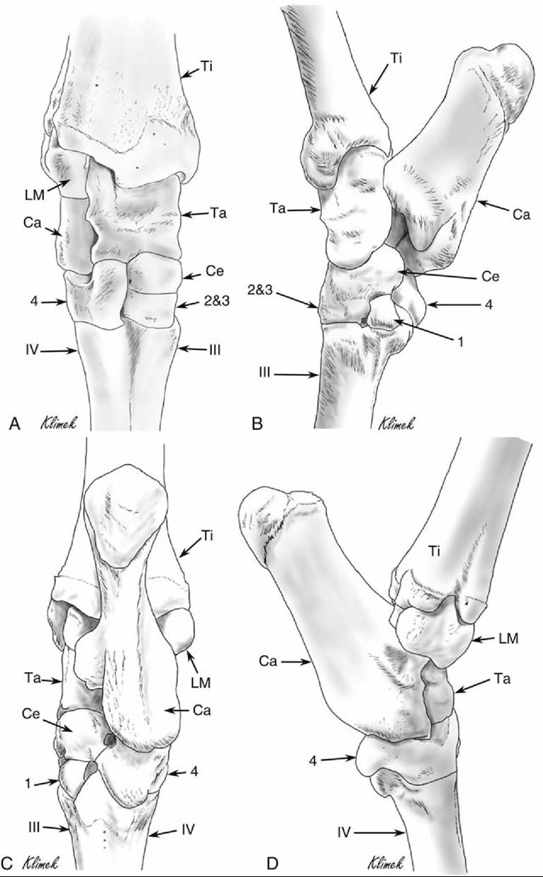
FIG. 38.33 Right tarsus of a llama. (A) Dorsal view. (B) Medial view. (C) Plantar view. (D) Lateral view. In this specimen, the central tarsal bone was partially fused with tarsal bone 2&3. Ca, Calcaneus; Ce, central tarsal bone; LM, lateral malleolus; Ta, talus; Ti, tibia; 1, first tarsal bone; 2&3, fused second and third tarsal bones; 4, fourth tarsal bone; III, third metatarsal bone; IV, fourth metatarsal bone.
There is wide individual variability in the extent that synovial joints communicate in the compound joints of the tarsus and carpus in the camelid. There are four separate synovial joints in
the tarsus. The tibiotarsal and proximal intertarsal joints always communicate; the proximal and distal intertarsal joints communicate at a rate of 34%; the distal intertarsal and tarsometatarsal joints communicate in 64% of cases; and in 26% of tarsi, all of the synovial joints communicate. There are differences between the right and left tarsus in 23% of animals.
The third and fourth metatarsal bones are fused as they are in other artiodactyls, with separate marrow cavities and separate articular surfaces at the distal end for each digit. The sagittal crest on each of these articular surfaces is only present on the plantar aspect of the articular surface. Each digit has two proximal sesamoids and the usual three phalanges, but there are no distal sesamoids in either the forelimb or the hindlimb digits. The bones of the digit are described more fully with the foot.
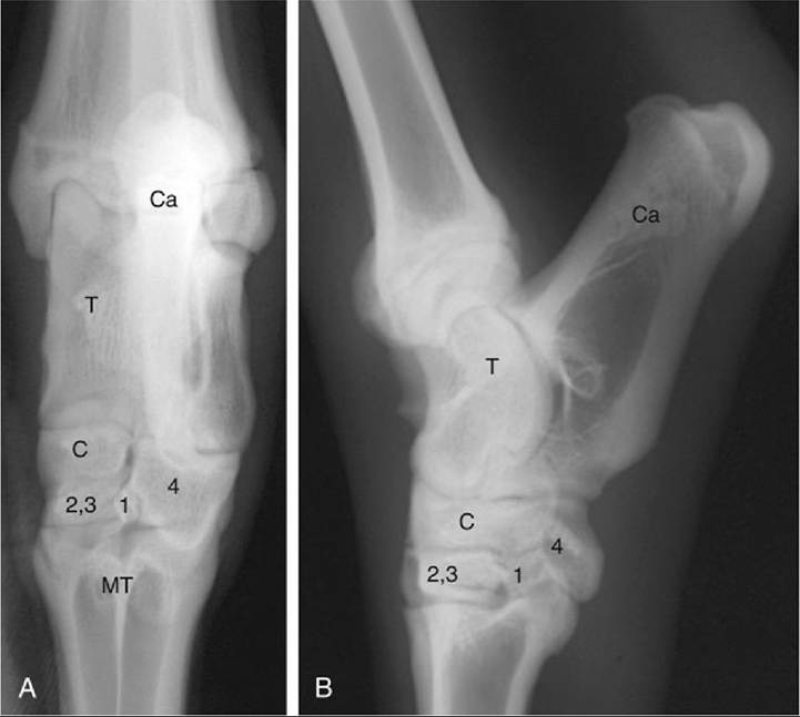
FIG. 38.34 Dorsopalmar (A) and lateral (B) radiographs of the llama tarsus. C, Central tarsal bone; Ca, calcaneus; MT, fused third and fourth metatarsal bones; T, talus; 1, first tarsal bone; 2,3, fused second and third tarsal bones; 4, fourth tarsal bone.
(From Cebra C, Anderson DE, Tibary A, et al: Llama and alpaca care: medicine, surgery, reproduction, nutrition, and herd health, St. Louis, 2014, Elsevier, Fig. 58-10.)Although there is not much specific information about the musculature of the llama or alpaca in the literature, the hindlimb digital flexor muscles and suspensory apparatus of the llama have been studied. Alpacas can be presumed to be similar. Proximal to the tarsus these muscles as well as the gastrocnemius are similar to those of ruminants. In the llama the soleus is absent. The deep digital flexor has three heads; lateral head, medial head, and caudal head. The tendons of the lateral head and the small caudal head join to travel together across the sustentaculum tali. The medial head crosses the medial malleolus and joins the main tendon distal to the sustentaculum tali. A small muscle not found in ruminants, the quadratus plantae, arises from the medioplantar aspect of the calcaneus, is deep to the flexor retinaculum, and inserts at the level of the fusion of the tendons of the deep digital flexor. There is no reciprocal apparatus, accessory ligament of the deep digital flexor tendon, or stay apparatus in the hindlimb of llamas.
Llamas have a pair of lumbricalis pedis muscles in the distal plantar aspect of the metatarsus, at the level of the bifurcation of the deep digital flexor tendon. The tendons of these muscles pass through the interdigital space and attach dorsally to the axial tendons arising from the lateral branch of the long digital extensor. Similar muscles are also present in the forelimb. Axial or abaxial extensor branches of the interosseus muscles are not present in llamas and alpacas. However, these lumbricalis pedis muscles (or lumbricalis manus muscles in the forelimb) have attachments similar to the axial extensor branches of the interosseus in ruminants.
The fetlock joints of the llama have ligaments that share characteristics with both the horse and the ruminants.
There is an interdigital metatarsointersesamoid ligament, which is present in the horse (as the metatarsointersesamoid ligament) but not in the ruminant. However, there is no straight sesamoidean ligament, a ligament found in horses. Llamas have oblique sesamoid ligaments, which are present in horses but not ruminants. However, instead of taking the equine form of the ligament, that of a solid sheet, llamas have separate axial and abaxial branches of this ligament. Interdigital phalangosesamoid ligaments of ruminants are replaced with interdigital metatarsophalangosesamoid ligaments, described for the first time in llamas. The axial proximal sesamoid bones are connected to each other via an interdigital intersesamoid ligament and to the distal end of the metatarsal bone via the interdigital metatarsointersesamoid ligament. Llamas additionally have short and cruciate sesamoid ligaments.Very little has been published in the English language regarding the vasculature and nerve supply of the pelvic limb of the llama or alpaca. The arteries and nonsatellite veins of the hindlimb in the llama have been described. In llamas there is a greater development and importance of the saphenous artery to the blood supply of the distal hindlimb than is typical of other domestic animals.
The main artery to the distal hindlimb in the llama is the caudal branch of the saphenous artery. This artery is accessible to take the pulse. It divides at the tuber calcanei into the medial and lateral plantar arteries, of which the medial is the larger and is the main source of blood to the distal limb. The cranial tibial and dorsal pedal arteries are small and not important to the blood supply of the hindfoot, and there is no perforating tarsal artery.
Among the more notable findings of the blood supply of the alpaca rear limb was the absence of a deep femoral artery in this species. Branches of the femoral artery generally follow the typical pattern, but the descending genicular artery originates from the popliteal artery rather than the femoral artery.
The branches of the cranial and caudal tibial and saphenous arteries distal to the tarsus are similar to those described in the llama.The distribution of the plantar arteries resembles that of the horse, but the medial vessels are much larger. The medial plantar artery gives off a deep branch at the base of the metatarsus and continues as the superficial branch between the deep digital flexor tendon and the medial plantar nerve. The superficial branch of the medial plantar artery moves axially at the level of the distal third of the metatarsus and, after anastomosing with plantar metatarsal artery III, gives rise to plantar common digital arteries II, III, and IV and distal perforating metatarsal artery III.
Plantar common digital artery III is much larger than the other two. It divides into axial plantar proper digital arteries III and IV; each of these gives off a plantar branch to the proximal phalanx that moves abaxially under the flexor tendons and then continues as the axial branch, supplying the dorsal artery of the middle phalanx, the coronal artery, and a branch to the digital pad, and ends in the dermal laminae. The plantar branch to the proximal phalanx continues as the abaxial plantar proper digital artery (III or IV), with branches similar to the axial plantar proper digital arteries. A terminal arch can be seen with contrast angiography.
One study found a superficial set and a deep set of veins in the pelvic limb of the llama. The medial saphenous vein is present. The deep set of veins generally follows the arteries, with some notable exceptions. There is a femoral vein and a medial circumflex femoral vein in the thigh, but the absence of the deep femoral artery as mentioned previously would imply that there is no medial circumflex femoral artery. There is also an extra vein in the thigh without a companion artery. This vein joins the femoral vein at the junction of the medial circumflex femoral and the femoral veins and appears to serve the area supplied by the lateral saphenous vein in other species.
It anastomoses with the popliteal vein distally. The popliteal vein is in the typical location. Coming off the caudal side of the popliteal vein is an expansion located between the heads of the gastrocnemius muscle, which may serve as a pump with contraction of the gastrocnemius muscle. This expansion is continued by a vein that ultimately anastomoses with the medial saphenous vein and may be analogous to the caudal branch of the medial saphenous vein of the horse. The medial saphenous vein connects to either the caudal tibial vein or the femoral vein directly. Although the lateral saphenous vein was reported to be absent in this investigation, other authors mention its use for venipuncture.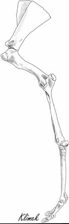
FIG. 38.35 Llama foreleg.
The nerve supply of the pelvic limb of the llama or alpaca has not been reported in the literature.