» Thoracic Limb
There is nothing remarkably different about the thoracic limb skeleton of the camelid compared to other livestock species. Fig. 38.35 is a drawing of the forelimb skeleton of the llama.
The spine of the scapula is unremarkable. It is offset such that the infraspinous fossa is larger than the supraspinous fossa. The coracoid process has a lip that angles medially. The humerus has an intermediate tubercle similar to that of the horse, with the greater and lesser tubercles being of equal size and both projecting proximal to the head of the humerus. The deltoid tuberosity is prominent. The ulna is partially fused to the radius, with its distal end visible as a distinct but fused portion and with remnants of the interosseus space both proximally and distally. The fused radius and ulna and the third and fourth metacarpal bones are very long and slender.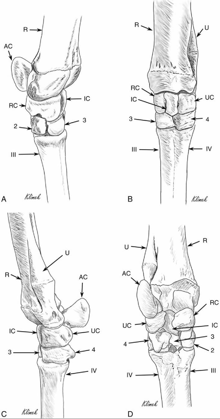
FIG. 38.36 Left carpus of a llama. (A) Dorsomedial view. (B) Dorsal view. (C) Lateral view. (D) Palmar view. AC, Accessory carpal bone; IC, intermediate carpal bone; R, radius; RC, radial carpal bone; U, ulna;
UC, ulnar carpal bone; 2, second carpal bone; 3, third carpal bone; 4, fourth carpal bone; III, third metacarpal bone; IV, fourth metacarpal bone.
The carpus has the accessory, radial, intermediate, and ulnar carpal bones in the proximal row and a small second carpal bone and larger and separate third and fourth carpal bones in the distal row. There are no sesamoids or first carpal bone in the llama. The individual joints can be palpated with the joint flexed, but there is a callus on the dorsal surface of the carpus that may make palpation more difficult. Fig. 38.36 shows the bones of the llama carpus, and Fig. 38.37 shows radiographs of the carpus of a llama.
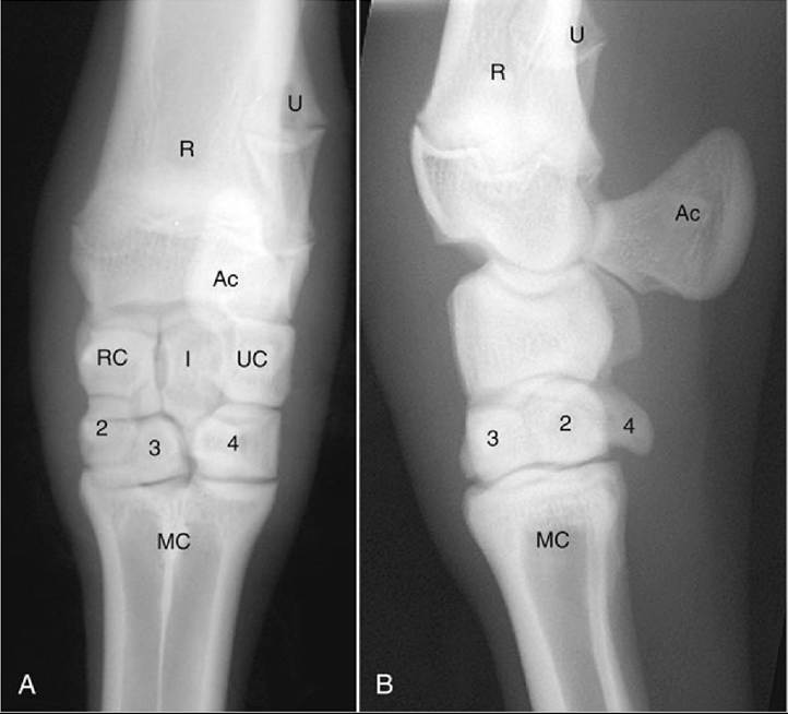
FIG.
38.37 Dorsopalmar (A) and lateral (B) radiographs of the llama carpus. Ac, Accessory carpal bone;I, intermediate carpal bone; MC, fused third and fourth metacarpal bones; R, radius; RC, radiocarpal bone; U, ulna; UC, ulnar carpal bone; 2, second carpal bone; 3, third carpal bone; 4, fourth carpal bone. (From
Cebra C, Anderson DE, Tibary A, et al: Llama and alpaca care: medicine, surgery, reproduction, nutrition, and herd health, St. Louis,
2014, Elsevier, Fig. 58-9.)
The carpal synovial joints communicate with each other with higher frequency than reported in horses and cattle. The radiocarpal joint communicates with the middle carpal joint in about one in three individuals, and the middle carpal joint always communicates with the carpometacarpal joint. The tendon sheaths for the carpus are typical of those in other species, but there is frequently (64%) communication between the carpal sheath (the tendon sheath for the superficial and deep digital flexors) and the radiocarpal joint in the llama.
The two metacarpal bones are fused, with features similar to those described for the hindlimb. The skeletal structures of the foot are described later.
The suspensory apparatus and digital flexor muscles of the forelimb have been described. The flexor carpi ulnaris was found to have a strong humeral head and a reduced ulnar head in the llama. In llamas there is only one belly of the superficial digital flexor muscle, in contrast to the two bellies that are found in ruminants. The superficial digital flexor tendon is relatively small and crosses the carpal canal in the superficial compartment. Distal to the carpus, the tendon of the superficial digital flexor joins the much larger palmar fascia; this is analogous to the accessory ligament of the superficial digital flexor as seen in the horse. This combined structure bifurcates at the fetlock into the two tendons serving the digits. The superficial digital flexor forms a manica flexoria in each digit, as in other species, and inserts on the middle phalanx via the middle scutum.
The deep digital flexor has the usual three heads—radial, ulnar, and humeral—and the humeral head additionally has a superficial and a deep portion. The tendons of all of these portions unite at the level of the carpus and are also joined by a small branch from the flexor carpi ulnaris muscle. The deep digital flexor bifurcates above the fetlock in the usual manner to pass to the distal phalanx of each digit. It is described in more detail later with the foot.
The interosseus muscle is mainly tendinous, as in the horse. It inserts via two symmetrical branches on the four proximal sesamoids, but it lacks the extensor branches found in horses and ruminants. The palmar fascia fuses on the lateral aspect with the origin of interosseous muscles III and IV (analogous to the suspensory ligament of the horse).
As is the case in ruminants, the common digital extensor (and the long digital extensor in the rear limb) has two tendons referred to as the medial and lateral tendons, and the lateral tendon divides to serve both digits. There is also a lateral digital extensor in both the forelimb and hindlimb. As described for the hindlimb, either one or two lumbricalis manus muscles are present on the palmar aspect of the metacarpus, at the level of the bifurcation of the deep digital flexor muscle; the number varies between individuals. These muscles, when present, each have a tendon that joins the axial extensor tendon of digit 3 or digit 4, a tendon that originates from the lateral head of the common digital extensor to serve either digit 3 or digit 4.
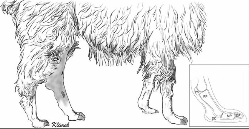
FIG. 38.38 Llama feet. Inset shows that camelids stand with the middle and distal phalanges parallel to the ground. There is no distal sesamoid. DC, Digital cushion; DP, distal phalanx; MP, middle phalanx; PP, proximal phalanx; PS, proximal sesamoid.
Little information about the specific pattern of vessel or nerve supply of the forelimb exists in the literature.