Placenta
The camelid placenta is chorioallantoic, diffuse, and epitheliochorial, with densely folded papillae on the chorionic surface. The ultrastructure of the papillae has been compared to the shape of a morel mushroom, with a constricted base and an expanded folded apex.
Other sources describe the chorionic surface as simply folded. The allantoic sac does not fill the space in the pregnant horn but extends into the full area of the nonpregnant horn. The amnion fills the space of the pregnant horn that is not occupied by the allantoic sac and is adhered to the inner surface of the chorion as well as to the allantois, which generally remains intact in unassisted deliveries. There is a normal 3-cm- wide nonvilliated area of the chorion along the lesser curvature, corresponding to the position of the main chorionic vessels. Hippomanes can be found in the allantoic cavity and are generally tan to dark brown in color, and amniotic plaques are normal. There is generally no remnant of the yolk sac. The umbilical cord of camelids has two umbilical arteries and, unlike most domestic animals, two umbilical veins and also contains the remnant of the allantoic stalk. The umbilical arteries and veins are similar in structure.Camelids have an extra fetal membrane derived from the epidermis and referred to as the epidermal membrane. It develops from the outer epidermis and adheres to the surface of the fetus until hair begins to form and pushes it away, after which some keratinization develops. In camels this separation from the surface of the fetus occurs in the latter fourth of gestation. The epidermal membrane covers the body of the fetus, being joined to it at mucocutaneous junctions and at the junction of the footpads and nails with the skin. It usually breaks down and wears off with little friction following birth. In camelids the amniotic fluid remains watery, lacking the mucus component that develops in the mare and cow, and the epidermal membrane may facilitate lubrication of the fetus.
Camelids do not lick their newborn or remove any fetal membranes.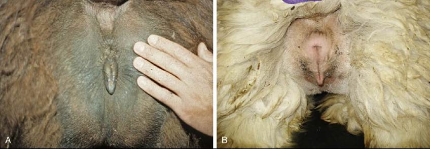
FIG. 38.30 Normal conformation of the vulva of the llama (A) and alpaca (B). Note the small perineal body and the prominent clitoris. (From Cebra C, Anderson DE, Tibary A, et al: Llama and alpaca care: medicine, surgery,
reproduction, nutrition, and herd health, St. Louis, 2014, Elsevier, Fig. 17-1.)
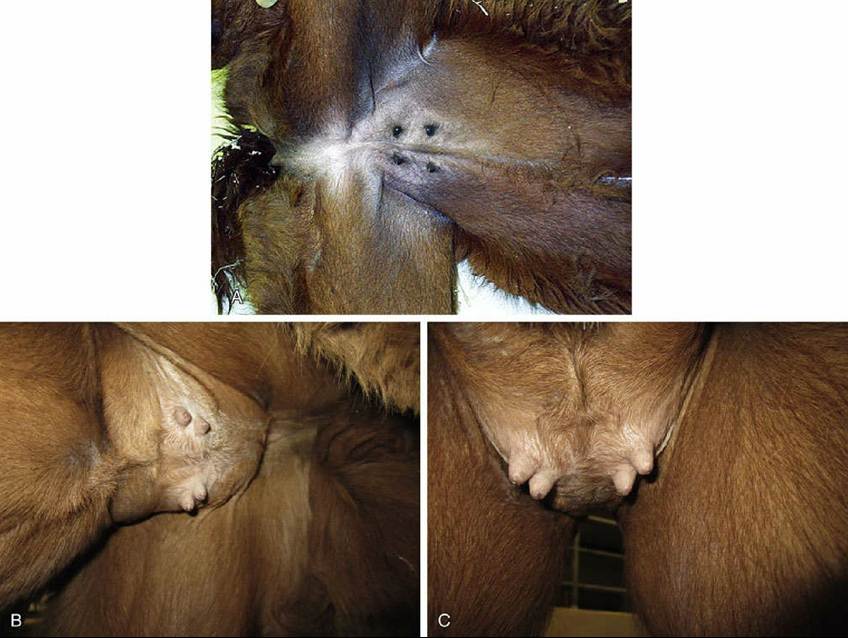
FIG. 38.31 Normal conformation of the udder in the nonlactating (A) and lactating (B and C) alpaca. (From
Cebra C, Anderson DE, Tibary A, et al: Llama and alpaca care: medicine, surgery, reproduction, nutrition, and herd health, St. Louis, 2014, Elsevier, Fig. 25-10.)
Delivered or Retained Placenta: The placenta is normally delivered within 1 h after birth. Parturitions that take place outside the normal parturition window are more likely to result in a retained placenta.