Placentation and Prenatal Development
In the horse, unlike other domestic species, a choriovitelline placenta (or omphalochorion) provides the principal organ of exchange for the first third or so of intrauterine life.
Thereafter, with the establishment of the chorioallantoic placenta, the yolk sac wanes. The definitive chorioallantoic placenta is of the epitheliochorial type and is commonly described as diffuse. The outer surface of the chorion carries innumerable branched villi that penetrate into crypts of the endometrial surface to form a loose attachment that is reinforced by the radial pressure exerted by the fetal fluids.Although the villi are widely spread, their distribution is not uniform, and they are clumped together in groups sometimes known as microcotyledons (because they resemble the cotyledonary arrangement in ruminants on a smaller scale). Small spaces between the microcotyledons face the openings of the uterine glands and fill with their secretions.
The capillaries of both fetal and maternal parts of the placenta reach directly below the corresponding epithelia, and only a thin tissue layer separates the two bloodstreams. Even so, the passage of large molecules, including antibodies, is impossible, and the passive transfer of immunity from mother to offspring is dependent on the foal ingesting colostrum.
A peculiar feature is the presence of so-called hippomanes in the allantoic (and, to a lesser extent, amniotic) fluid. Most of these soft brownish bodies are formed by the deposition of mucoproteins and calcium phosphate on nuclei provided by solid particles within the fluids, but some originate in material flaked from endometrial cups when these have completed their role. The latter are sometimes found anchored to the chorioallantoic membrane by attenuated stalks. Hippomanes have no known clinical (or residual physiologic) importance.
Although detailed information must be sought elsewhere, it may be useful to have a basic guide to the estimation of fetal age (Table 22.1). Crown-rump measurements are of limited value in this species because of its wide range of body size.
» TABLE 22.1
Guide to the Aging of Horse Fetuses
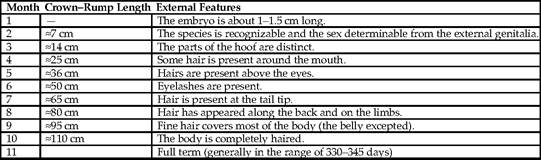
From Evans HE, Sack WO: Prenatal development of domestic and laboratory animals. Growth curves, external features and selected references, Anat Histol Embryol 2:11—45, 1973.
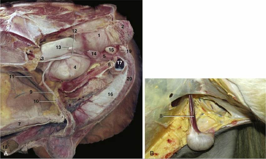
FIG. 22.19 (A) The reproductive organs of the stallion in situ. 1, Rectum; 2, external anal sphincter; 3, ureter; 4, bladder; 5, urethra; 6, floor of pelvis; 7, floor of abdomen; 9, left deferent duct; 10, vaginal ring;
11, right testicular artery and vein; 12, ampulla of deferent duct; 13, vesicular gland; 14, prostate; 15, bulbourethral gland; 16, penis; 17, left crus (in section); 18, glans penis; 19, ischiocavernosus; 20,