RESPIRATORY APPARATUS
1. Define and locate visceral pleura and costal pleura.
2. What is the mediastinal space?
3. What structures lie within the mediastinal space?
4. What happens to mediastinal pressure when intrapleural pressure decreases?
5.
How are the nostrils of the horse adapted to the need for greater air intake?6. What functions are served by the conchae?
7. Where is the olfactory epithelium located?
8. List the openings to the pharynx.
9. What is the function of the pharynx and syrinx?
0. What is the function of tracheal rings? Why are they incomplete dorsally?
11. What are the subdivisions of the trachea (in order from largest to smallest)?
2. Where does most of the diffusion of gas between air and blood occur?
The respiratory apparatus consists of the lungs and pleura and the air passages leading to the lungs, including the nostrils, nasal cavities, pharynx, larynx, trachea, bronchi, and bronchioles.
Airways to the Lungs
The nostrils (nares) are the paired external openings to the air passages (Figure 10-1). The nostrils are the most pliable and dilatable in the horse and the most rigid in the pig. Nostril dilatation is advantageous when more air is required, as in running, and in situations in which breathing is not done through the mouth. The horse is a runner and open-mouth breathing is not characteristic; therefore dilatable nostrils are advantageous.
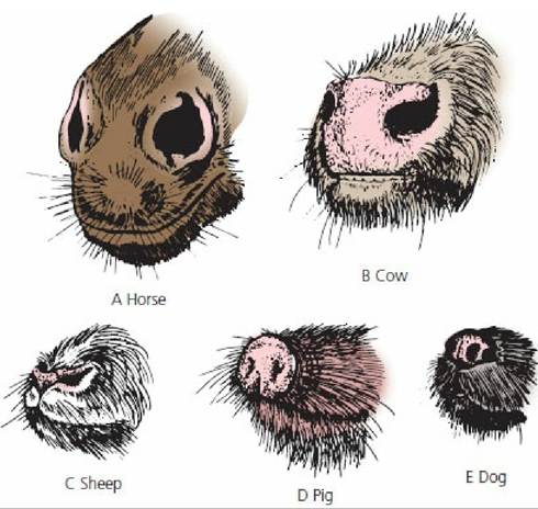
■ FIGURE 10-1 The nostrils of several domestic animals. A. Horse. B. Cow. C. Sheep. D. Pig. E. Dog. (From Frandson RD, Wilke WL, Fails AD. Anatomy and Physiology of Farm Animals. 7th edn. Ames, IA: Wiley-Blackwell, 2009.)
The nostrils provide the external openings for the paired nasal cavities. The nasal cavities are separated from each other by the nasal septum and from the mouth by the hard and soft palates.
In addition, each nasal cavity contains mucosa-covered turbinate bones (conchae) that project to the interior from the dorsal and lateral walls, separating the cavity into passages known as the common, dorsal, middle, and ventral meatus (Figure 10-2). The mucosa of the turbinates is well vascularized and serves to warm and humidify inhaled air. Another function, mainly of the conchae, that is often overlooked involves cooling the arterial blood that supplies the brain. Arteries that supply blood to the brain divide into many smaller arteries at its base and then rejoin before entering. These smaller arteries are bathed in a pool of venous blood that comes from the walls of the nasal passages, where it has been cooled. As a result, brain temperature might be 2 or 3 °C lower than the body core temperature. The brain is the most heat-sensitive body organ, so this cooling method is particularly important during times of extreme activity. The mouth breathing that occurs when the environmental air is extremely cold seems to be reflexive, which might prevent the overcooling of the brain that could otherwise occur if all of the inhaled air traversed the meatus and had contact with the conchae. The olfactory epithelium is located in the caudal portion of each nasal cavity and greater perception of odors (a nonrespiratory function) is achieved by sniffing (i.e., fast, alternating, and shallow inspirations and expirations).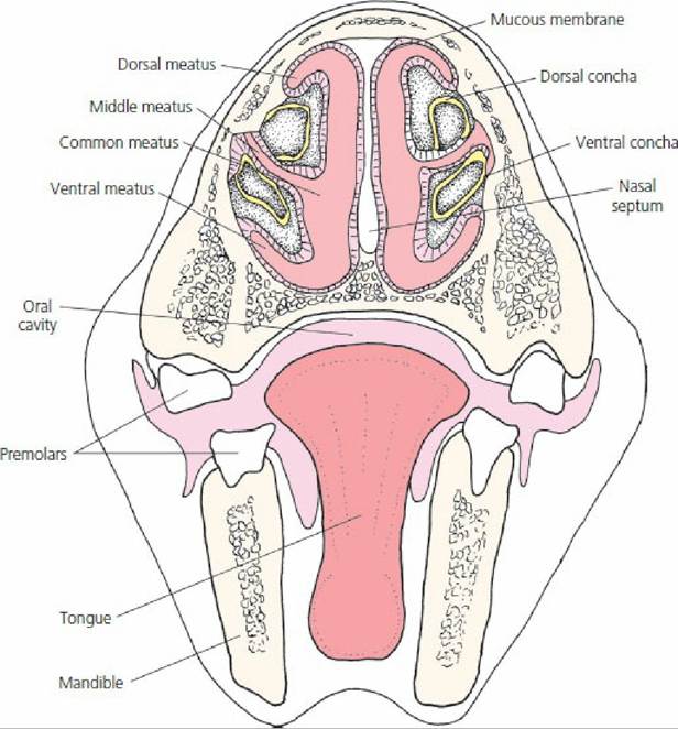
■ FIGURE 10-2 Transverse section of the head of a horse showing the division of the nasal cavities. The airways are noted as the dorsal, middle, ventral, and common meatus. The conchae consist of turbinate bones covered by a highly vascularized mucous membrane. It can be seen that incoming air is exposed to a large surface area for adjustment of its temperature and humidity.
The pharynx is caudal to the nasal cavities and is a common passageway for air and food (Figure 103).
The openings to the pharynx include two posterior openings from the nasal cavity (choanae), two eustachian tubes, a mouth (oral cavity), a glottis, and an esophagus. The opening from the pharynx leading to the continuation of the respiratory passageway is the larynx, the organ of phonation (sound production) in mammals. Sound is produced by the controlled passage of air, which causes vibration of vocal cords in the larynx. The organ of phonation in birds is called the syrinx; this is located where the trachea divides to form the bronchi.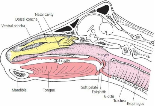
■ FIGURE 10-3 Midsagittal section of the head of a cow with the nasal septum removed. The stippled area represents the pathway for air through the nasal cavity, pharynx, larynx, and trachea. The glottis is the opening to the larynx that is continued caudally by the trachea.
The glottis is the slit-like opening between the vocal cords and is the site for insertion of an endotracheal (within the trachea) tube when it is used for providing assisted ventilation and for administration of inhalant anesthetics. Extending the cranium from the larynx is the epiglottis. It is a leaf-shaped plate of cartilage covered with a mucous membrane that is located at the root of the tongue and is passively bent over the larynx during the act of swallowing, thereby preventing the entrance into the trachea of a bolus of food. A cranial view of the glottis and epiglottis as they would appear with the mouth open and the tongue extended is shown in Figure 10-4. In this view, the soft palate (caudal extension of the hard palate) has been hyperextended with the maxilla (upper jaw). When placing an endotracheal tube, the soft palate is often seen ventral to the epiglottis with usual mouth opening and must be lifted by manipulation of the endotracheal tube to expose the glottis. Figure 10-5 shows an endotracheal tube in place relative to the structures encountered.
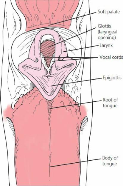
■ FIGURE 10-4 Cranial view of canine glottis (opening to the larynx, between the vocal cords) and epiglottis (cranial extension from the larynx).
The soft palate is not shown in the location that would be seen with usual mouth opening techniques.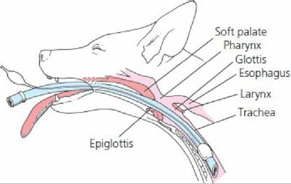
■ FIGURE 10-5 Schematic view of an endotracheal tube in place relative to the structures encountered.
The trachea is the primary passageway for air to the lungs. It is continued from the larynx cranially and divides caudally to form the right and left bronchi. The tracheal wall contains cartilaginous rings to prevent collapse of the tracheal airway (Figure 10-6). Each tracheal ring is incomplete (not joined dorsally), which permits variations in diameter that are regulated by the tracheal smooth muscle. This diameter can increase during times of greater ventilatory requirements.
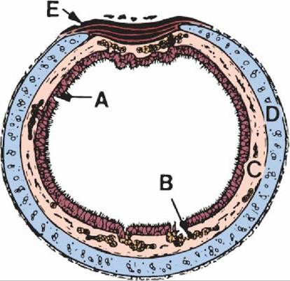
■ FIGURE 10-6 Schematic representation of a cross-section of trachea. A. Pseudostratified epithelium lines the lumen. B. Glands in the lamina propria. C. Glands in the submucosa. D. Cartilage. E. Band of smooth muscle. The tracheal muscle and the cartilage form most of the tracheal wall. (From Eurell JA, Frappier BL. Dellmann’s Textbook of Veterinary Histology. 6th edn. Ames, IA: Blackwell Publishing, 2006.)
The right and left bronchi and their subdivisions continue all the way to the alveoli, the final and smallest subdivisions of the air passages (Figure 10-7). The subdivisions of the trachea to the alveoli, from the largest to the smallest, comprise bronchi, bronchioles, terminal bronchioles, respiratory bronchioles, alveolar duct, alveolar sac, and alveoli.
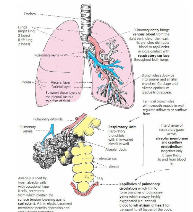
■ FIGURE 10-7 Schematic representation of lung subdivisions. (From Mackenna BR, Callander R. Illustrated Physiology. 6th edn. Edinburgh: Churchill Livingstone, 1997.)
Pulmonary Alveoli
The pulmonary alveoli are the principal sites of gas diffusion between the air and blood.
The separation of air and blood, and thus the diffusion distance, is minimal at the alveolar level. The alveolar epithelium and the capillary endothelium are intimately associated (Figure 10-8). Here, venous blood from the pulmonary arteries becomes arterial blood and is returned to the left atrium by the pulmonary veins. The darker purple color of venous blood becomes bright red arterial blood during the resaturation of hemoglobin with new oxygen that has diffused from the alveoli. During the seventeenth century, Richard Lower showed that the change in blood color occurred in the lungs because of the influence of fresh air. The idea that the diffusion of oxygen and carbon dioxide between blood and air was separate from a secretion process was proven by August and Marie Krogh. (August Krogh won the Nobel prize in 1920 for his studies of the capillaries.)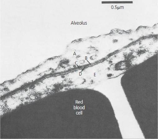
■ FIGURE 10-8 Electron micrograph of a mouse lung showing an attenuated portion of alveolar epithelium and its proximity to capillary endothelium. The respiratory membrane (without alveolar fluid layer) is composed of the following: A. alveolar epithelium, B. alveolar epithelial basement membrane, C. interstitial space, D. capillary endothelial basement membrane, and E. capillary endothelium. (From Reece WO. Respiration in mammals. In: Reece WO, ed. Dukes’ Physiology of Domestic Animals. 13th edn. Ames, IA: Wiley-Blackwell, 2015.)
The Lungs and Pleura
The lungs are the principal structures of the respiratory system. They are paired structures and occupy all space in the thorax that is not otherwise filled. When the thorax expands in volume, the lungs also expand; this provides for air flow into the lungs. Air is an excellent radiographic contrast media because it is radiolucent (relatively penetrable by X-rays). Therefore, air-filled lungs provide a good contrast for thoracic structures (normal and pathologic) that are radiopaque (relatively impenetrable by X-rays).
Dorsal-ventral and lateral view radiographs of a normal canine thorax are shown in Figure 10-Q. The radiopaque objects (heart and blood vessels) appear superimposed on the radiolucent background of air. The heart and blood vessels are visible because the blood contained within is relatively radiopaque. The blood vessels appear as branching white tubes.
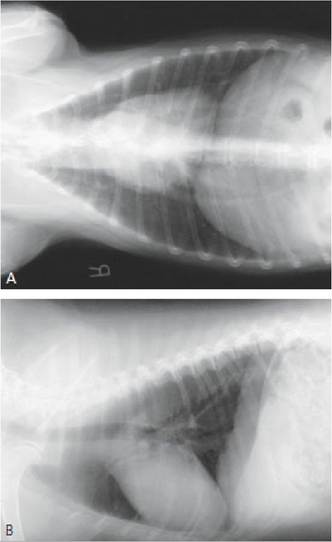
■ FIGURE 10-9 Radiographs of healthy canine thorax. A. Dorsal-ventral view. Radiographs of healthy canine thorax. B. Lateral view. The heart and major blood vessels are visible because blood is relatively radiopaque. Blood in lesser blood vessels gives a slightly cloudy appearance to the lung field as compared with the clear appearance of air in the trachea. (Radiographs courtesy of Dr Elizabeth Riedesel, Lloyd Veterinary Medical Center, Radiology Section, Iowa State University.)
The lungs have an almost friction-free movement within the thorax because of the pleura, a smooth serous membrane. The pleura is composed of a single layer of cells fused to the surface of a connective tissue layer covering the thoracic cavity. The pleural covering creates two pleural cavities (also referred to as intrapleural space), one on the right and one on the left sides. In normal individuals the pleural cavity is best thought of as a potential space and only contains a small amount of fluid. Only in certain conditions where air or fluid (blood, pus, etc.) is introduced into the cavity can it be fully appreciated. The pleura is referred to as parietal (covering the walls) or visceral (covering the lung). The parietal pleura is further subdivided into mediastinal, costal, or diaphragmatic depending on what structure it is covering.
The area near the midline in between the pleural cavities is mediastinal space. The mediastinum contains the venae cavae, thoracic lymph duct, esophagus, aorta, arteries and veins for the head and thoracic limb, lymph nodes, and trachea (Figure 10-10) Pressure changes in the pleural cavities (intrapleural space) are associated with pressure changes in the mediastinal space. Also, pressure changes within the mediastinal space are accompanied by changes within the mediastinal structures, provided that their walls are responsive to relatively low-pressure distensibility.
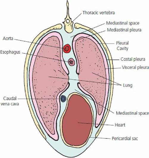
■ FIGURE 10-10 Schematic transverse section of equine thorax showing the relationships of the visceral, costal, and mediastinal pleura. The aorta, esophagus, venae cavae, and thoracic lymph duct (not shown) are within the mediastinal space. The esophagus, venae cavae, and lymph duct (soft structures) respond by increasing and decreasing pressures within their lumens, associated with similar changes in intrapleural and mediastinal spaces.
■