Respiratory Rhythmicity Originates in the Medulla and Is Modified by Higher Brain Centers and Inputs from Peripheral Receptors
Some of the early attempts to understand the brain’s role in the regulation of breathing involved experiments in brainstem transection to identify the regions of brain involved in maintenance of rhythmic breathing.
Fransection between the spinal cord and medulla arrests breathing, which indicates that the rhythnιicity Ofbreathing originates in the brain, not in the spinal cord or respiratory muscles. The effects of sectioning at different levels of the midbrain vary depending on whether the vagus is intact or cut. The results of these types of experiments have been interpreted as showing that respiratory rhythmicity originates in the medulla but is fine-tuned by afferent information received from the vagus and by higher centers in the brain, most importantly, the pons.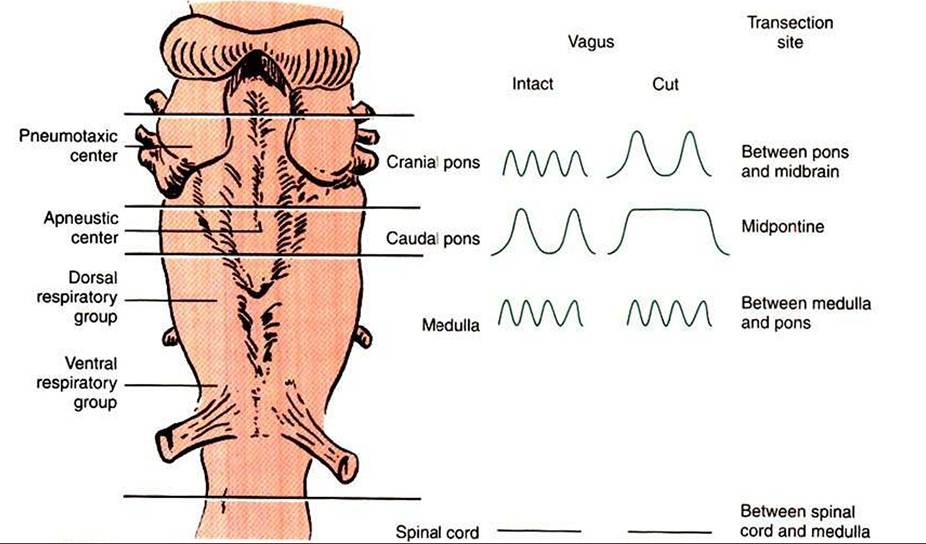
FIGURE 49-2 Ventral surface of the brainstem, including the pons, medulla, and spinal cord, showing centers involved in the regulation of respiration. The effect of sectioning the brain at the levels designated by the horizontal lines with the vagus intact or cut is shown in the recordings of breathing pattern on the right side of the diagram.
More recently, electrical recordings have identified three groups of neurons in the pons and medulla that fire synchronously with breathing (Figure 49-2). The pontine respiratory group (also known as the pneumotaxic center) lies in the dorsal lateral pons, receives input from vagal reflexes related to lung volume, and modulates respiratory frequency. Within the medulla, two groups of neurons fire in association with res- piration. The dorsal respiratory group is located in the ventral lateral portion of the nucleus tractus solitarius. The ventral respiratory group is located in the nucleus ambiguus and retroambiguus. The neurons of the dorsal respiratory group neurons fire primarily during inhalation, and those of the ventral respiratory group fire during both inhalation and exhalation.
The axons from the dorsal respiratory group project through bulbospinal pathways to inspiratory spinal motoneurons (primarily those supplying the diaphragm) and to the ventral respiratory group. Axons from the ventral respiratory group project to spinal motoneurons of both expiratory muscles and accessory inspiratory muscles.The origin of rhythmic breathing is currently unknown. Pacemaker neurons have been identified in the pre-Bδlzinger complex of the ventral respiratory group, but their role in normal breathing is unclear. Normal respiration (eupnea) seems to result from rhythmic inhibition of inspiratory activity. During inhalation there is an increase in activity in the inspiratory neurons. This increased activity is further amplified by an increase in the chemical respiratory drive, such as hypoxia. Termination Ofinspiration can be a result of vagal inputs from pulmonary stretch receptors or from a central pontine “off” switch. After vagotomy, the pontine “off” switch terminates inhalation after a fixed time for inhalation, which is independent of chemical drive. When the vagus is intact, and thus signals from pulmonary stretch receptors are relayed to the brain, there is a complex interaction between the time for inhalation and the tidal volume. This interaction leads to a larger tidal volume and more rapid respiratory frequency when the chemical drive to breathe is increased.
When inhalation is terminated, inspiratory neurons are inhibited, and thus exhalation occurs passively as a result of the elastic recoil of the lung and chest wall. Activity in some inspiratory neurons early in exhalation leads to inspiratory muscle activity, which provides a “brake” on exhalation and regulates the rate of expiratory airflow. Later in exhalation, the braking is removed. During this latter part of exhalation, expiratory muscles may be activated. When respiratory drive is low, this second phase of exhalation is initiated later than when drive is increased.
The “automatic” breathing just described is frequently overridden by demands from higher brain centers. Vocalization, parturition, swallowing, defecation, and many other activities require the active participation of the respiratory system.