Senior management: Brainstem and motor cortex
Key points
■ The majority of UMN nuclei in quadrupeds are located in the brainstem; however skilled movements are controlled by UMNs mainly located in the forebrain.
■ UMN originating in the motor cortex travel to the brainstem and synapse on LMNs in cranial nerve nuclei (corticonuclear tract).
Or they continue caudally via the pyramids of the medulla oblongata to synapse on LMNs in the spinal cord (corticospinal or pyramidal tract).■ Nuclei associated with maintaining posture (antigravity support) are the vestibular and pontine reticular formation. The red nucleus and medullary reticular nuclei facilitate flexor activity.
■ UMNs in the caudal brainstem that ultimately inhibit extensor muscle activity require input from midbrain or forebrain to function: facilitators of extension do not. Thus animals with lesions in the rostral brainstem can show increased extensor tone of the limbs and body (opisthotonus).
■ Senior management of the sensory system includes the cerebellum, which receives subconscious proprioceptive information, and specific receiving areas in the forebrain for conscious perception.
UMNs and the brain
Senior management of the motor system comprises UMN nuclei located primarily in the brainstem and the motor cortex of the cerebrum (Fig. 4.11). It makes feedback loops with the cerebellum. It talks with junior management (interneurons) in the brainstem and spinal cord and may communicate directly with the worker LMNs. The UMN system is managed by the executive officers located mainly in the forebrain.
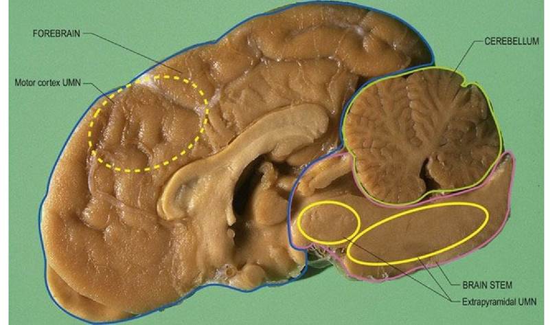
Fig. 4.11 Dog brain median section depicting location of UMN centres and nuclei in yellow outline. Note the motor cortex (dotted outline) in the forebrain is located primarily on the lateral aspect of the hemisphere with some extension over the medial edge into the brain bordering longitudinal fissure.
The senior management UMN system located in the forebrain is the motor cortex. It is organised somatopically such that specific regions of the cortex relate to specific regions of the body (see Fig. 4.14 for motor homunculi). Its output links to LMNs of the brainstem and the spinal cord via the corticonuclear (old name = corticobulbar) and corticospinal tracts, respectively.
The output from the motor cortex travels via the internal capsule in the forebrain and the crus cerebri into brainstem forming the longitudinal fibres of the pons. The corticonuclear fibres terminate on the cranial nerve nuclei in the brainstem. (Termination is primarily ipsilateral (CN IV) bilateral (CNN III, V, VII, IXXI) or contralateral (CNN VI, XII).) The remaining corticospinal tracts travel in the superficially located pyramids on the ventral aspect of the medulla oblongata forming the pyramidal system (see Chapter 5). In the spinal cord, the corticospinal tract divides into the lateral and ventral tracts, travelling caudally in the relevant funiculi. These tracts decussate, thus the motor cortex influences the contralateral side of the body. Decussation is either at C1 level (lateral corticospinal tract) or just prior to terminating (ventral corticospinal tract). The motor cortex and its outputs are much more important in primates, and especially in humans, than in domestic mammals. For example, in equids the corticospinal tract only extends as far as the second cervical spinal cord segment. An animal’s ability to perform skilled, dextrous movements is directly related to the development of the motor cortex and the pyramidal system (see Fig. 5.4).
In quadrupeds, the main UMN nuclei are found throughout the brain, but particularly in the brainstem. The UMN tracts arising from brainstem nuclei do not travel through the medullary pyramids and are thus known as the extrapyramidal system.
In the midbrain, UMN nuclei include the red nucleus (in the ventral midbrain, tegmentum) and tracts arising from the tectum (dorsal midbrain).
The pons is associated with the pontine reticular formation and the medulla oblongata with the medial and lateral medullary reticular formation and the vestibular nuclei. The locations and functions of the tracts arising from these UMN centres are given in Fig. 4.5 and Table 4.3. In summary, brainstem nuclei associated with maintaining posture (antigravity support) are in the vestibular and pontine reticular formation, while UMN centres associated with facilitating flexor activity/protraction are the red nucleus, medullary reticular formation and the motor cortex of the forebrain.Executive management centres located throughout the cerebral cortex activate the UMN system (Fig. 4.12).
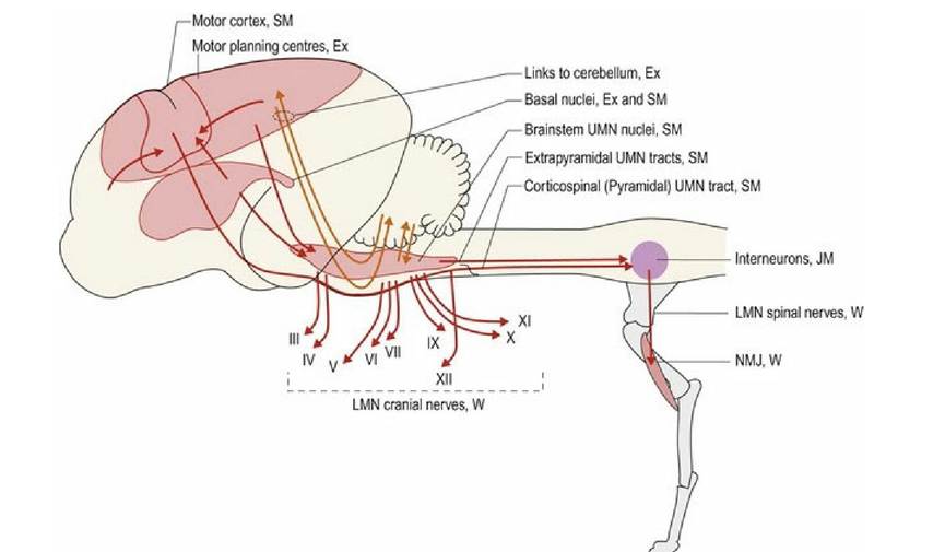
Fig. 4.12 Motor system hierarchy in the quadrupedal mammalian brain, including the motor planning centres of the executive management (see next section). Ex = motor executive, SM = senior management, JM
= junior management, W = worker.
Many of the UMN pathways facilitate movement, but some, especially the medial medullary reticulospinal tract, are major inhibitors of movement. However, the inhibitory UMNs, particularly those in the pons and medulla oblongata, require input from centres in the midbrain or forebrain to function - facilitators of extension do not. Thus an animal with a caudal fossa lesion, i.e. a lesion in the caudal part of the cranial vault, disrupting connections between the forebrain and caudal brainstem can show greatly exaggerated, generalised, extensor tone called ‘opisthotonus’ (see Fig. 9.6). Extensor (antigravity) muscle activity is stronger than flexor muscle activity. Therefore, in spinal cord lesions that damage UMN tracts, loss of LMN inhibition by the medial medullary reticulospinal tract, and loss of input from other tracts that facilitate flexor activity (e.g. corticospinal and rubrospinal), can result in significant spasticity, hypertonus and uninhibited reflexes (Fig.
4.2).All of these UMN centres have bidirectional links with the cerebellum, forming feedback circuits. Thus the UMN centre informs the cerebellum of its intended activity and the cerebellum then can regulate the movement as it occurs. To do this the cerebellum uses continuous subconscious proprioceptive feedback from the parts of the body that are moving and those that are involved in postural support (see Chapters 6 and 7).
Sensory systems
Senior management of the sensory systems comprises the primary receiving areas for incoming information, which are located in the cerebellum and forebrain (Fig. 4.13). The cerebellum receives subconscious proprioceptive information from muscle spindles and Golgi tendon organs of the body and limbs via the PNS and spinal cord. Subconscious proprioception for the head, originating in the vestibular apparatus of the inner ear, is sent to vestibular nuclei of the brainstem and hence to the cerebellum. Proprioceptive information from the head musculature is forwarded from the trigeminal nuclei in the brainstem. Conscious proprioceptive, tactile, nociceptive, thermal and gustatory information is received in the somatosensory cortex of the forebrain (see Fig. 4.14 for human and feline sensory homunculi). Auditory, visual and olfactory stimuli project to specific receiving areas in the forebrain, such as auditory and visual cortices and the olfactory lobe (Fig. 4.13).
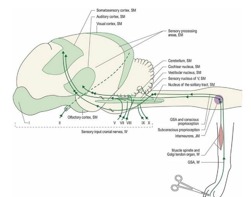
Fig. 4.13 Sensory system hierarchy in the quadrupedal mammalian brain. Roman numerals = cranial nerves, GSA = general somatic afferent, which includes tactile, thermal and nociception. Note the executive components (association and interpretation areas are not indicated on this figure, see next section).
Both the motor cortex and somatosensory cortex are somatopically arranged, such that the body areas can be mapped to specific areas of each cortex. This mapping results in the homunculus image (L = little man) (Fig. 4.14A), or in the case of the cat, the felunculus (Fig. 4.14B).
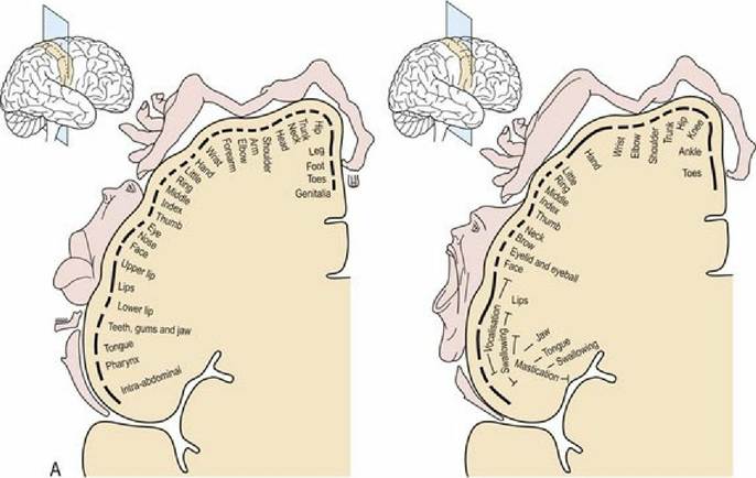
(redrawn with permission from Gray’s Anatomy 39th edition, fig 22-13).
Fig. 4.14A The human sensory (left) and motor (right) homunculus
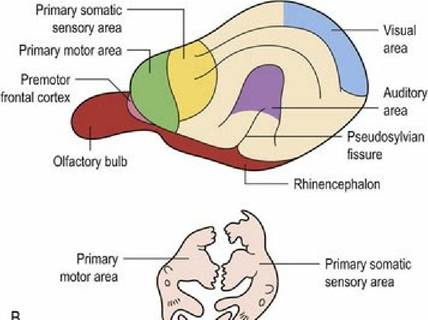
Fig. 4.14B The feline sensory and motor homunculi (redrawn with permission from King A.S. Physiological and Clinical Anatomy of the Domestic Mammals. Volume 1, Central Nervous System. Oxford University Press, 1993, figure 8.2b, page 102). The two figures are drawn at the same scale. The lower image of the homunculi can be superimposed on the primary motor and sensory areas, by spreading
them over both the medial and lateral aspects of the hemispheres.