SlowWaves of Electrical Depolarization Are a Unique Feature of Gut Smooth Muscle
The first level of control of Gl motility lies in the intrinsic electrical properties of the smooth muscle mass. These electrical properties consist of spontaneously undulating waves of partial depolarization that sweep over the gut smooth muscle.
The origin of this electrical activity is from specialized smooth muscle cells referred to as the interstitial cells of Cajal (ICC). The ICC form an interconnecting lattice of cells that surrounds the circular and longitudinal layers of muscle over the entire length of the gut. These cells are very similar in structure and function to the Purkinje cells of the heart. The ICC exhibit rhythmical and spontaneous oscillation in their transmembrane electrical potentials, as illustrated in Figure 28-1. They are connected to one another and to cells of the general smooth muscle mass by tight junctions or nexuses. These connections allow for the flow of ions from cell to cell. The resulting ionic movements lead to the propagation of waves of partial cell membrane depolarization across large numbers of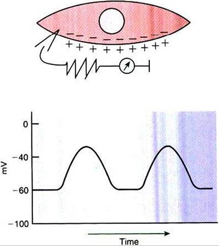
FIGURE 28-1 Spontaneous changes in membrane polarity of the interstitial cells of Cajal, specialized gastrointestinal (GD smooth muscle cells that are responsible for spontaneous electrical rhythmicity in gut muscle.The upper illustration represents a single cell with a voltmeter measuring the transmembrane electrical potential.The graph illustrates spontaneous changes in electrical potential (in millivolts, mV} that would be measured across the cell membrane.
cells. Within the ICC, fluctuations in intracellular calcium concentrations appear responsible for the spontaneous changes in membrane polarization. Figure 28-1 illustrates the concept of a fluctuating membrane potential in a single ICC. The property of spontaneous electrical rhythmicity, in combination with their electrical connection to the smooth muscle mass, imparts to the ICC their role as electrical “pacemakers” of the gut.
In GI smooth muscle cells the baseline membrane potential is usually-70 to -60 millivolts (mV). Under the influence of the ICC, the membrane potential fluctuates from this baseline level by as much as 20 to 30 mV. Thus, under resting conditions, the depolarization is only partial, and the membrane potential never reaches 0 mV. The smooth muscle cells are connected to the ICC and to each other by nexuses, allowing the changes in membrane potential to be spread, or propagated, over large areas of muscle. The ICC initiate these changes and thus determine their origin and direction of propagation. Under normal conditions in the small intestine, changes in membrane potential begin high in the duodenum and are propagated aborally (away from the mouth) along the length of the small intestine (Figure 28-2). These aborally moving waves of partial depolarization are called slow waves or the basic electrical rhythm of the gut. In the dog, slow waves Occurabout 20 times per minute in the small intestine. In the stomach and colon, slow waves occur less frequently, about five times per minute. However, the slow waves are present
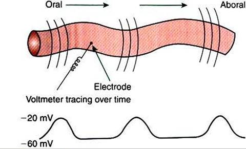
FIGURE 28-2 ■ Partial membrane depolarizations of Gl smooth muscle cells occur in a coordinated manner, creating waves of depolarization that sweep over large segments of muscle. Electrodes placed on or near the surface of the muscle record changes in potential as waves of depolarization pass toward or away from them. Coordinated changes in membrane potential among cells are necessary for these waves to be measured because random changes among cells would cancel each other out, and no changes would be recorded by electrodes placed e?tracellularly.
throughout the smooth muscle portions of the GI tract. The frequency of slow waves varies among the domestic species, but their presence does not vary.
The slow waves are an intrinsic property of the GI smooth muscle and associated ICC. The presence of the slow waves depends only on the ICC, whereas the amplitude and to a lesser extent the frequency of the slow waves can be modulated by the enteric nervous system (ENS) and endocrine/paracrine system. The link between slow waves and muscle contractions, however, is under control of nervous, endocrine, and paracrine factors, as discussed next.