SOMATIC MOTOR PATHWAYS
Somatic motor activity is regulated at two levels within the central nervous system by separate groups of nerve cells conveniently designated the lower and upper motor neurons.
The lower motor neurons are located within the ventral column of the gray substance of the spinal cord (Figure 8-12Z√) and within the somatic motor nuclei of those cranial nerves that contain somatic efferent components.
Their axons are conveyed within the spinal and relevant cranial nerves to the skeletal muscles, where each terminates on a group of muscle fibers (Figure 8-48); the size of the group varies with the precision of performance required of the particular muscle (p. 25). Lower motor neurons provide the efferent limbs of simple reflexes but are in most other circumstances directed by upper motor neurons.The upper motor neurons are involved in more complicated reflexes and also initiate voluntary movements. They are mainly located within the motor area of the neopallium but also in other regions of the brain, including the reticular formation and red nucleus. The cortical areas allocated to neurons controlling the muscles of different parts of the body vary in extent with the importance and complexity of the movements of these parts in the habitual activities of the species; thus the hand occupies a relatively much larger area of the human cortex than that allocated to the whole limb in ungulates. Upper motor neurons do not project
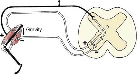
Figure 8-48 A myotactic reflex arc. Gravity (arrow)stretches the extensor muscle, stimulating its contraction via the reflex arc. The flexor muscle is inhibited by a collateral fiber and an inhibiting interneuron.
directly on muscle fibers but exert their control by excitation or inhibition of lower motor neurons.
The connections of the upper with lower motor neurons follow various pathways that vary considerably among species in their relative development and details of organization. The primary distinction is made between so-called pyramidal and extrapyramidal systems, although the two are coordinated and work in close collaboration. The pyramidal system is mostly concerned with the exercise of finely adjusted movements, while the extrapyramidal system is employed in the control of coarser movements, particularly in stereotyped locomotor patterns. It follows that the pyramidal system must be better developed in primates than in domestic species, which is a distinction that explains the different consequences of lesions to the pyramidal pathway. Severe damage to the pyramidal pathway produces a complete and permanent paralysis of the contralateral voluntary musculature in ourselves, while the effects in domestic species are mainly confined to disturbance of contralateral postural reactions from which partial recovery occurs after a few days. Both pyramidal and extrapyramidal systems are provided with elaborate feedback mechanisms that allow for the continuous monitoring and adjustment of motor activity.
The Pyramidal System
The pyramidal system takes origin from neurons within various regions of the neopallium, particularly the primary motor area. The axons of these neurons converge toward their exit from the telencephalon and form an important fraction of the internal capsule; in their passage they preserve the orderly point-to-point arrangement of the cortical representation. They then continue over the lateral aspect of the thalamus to enter the crus cerebri on the ventral surface of the brain (Figure 8-33Z9); after traversing the ventral portion of the pons, they reappear on the surface as the pyramids of the medulla oblongata (Figure 8-19Z17). Three fiber groups may be distinguished within the system: corticospinal fibers continue through the medulla oblongata into the spinal cord; corticobulbar fibers peel off at appropriate levels of the brainstem to reach various nuclei of contralateral cranial nerves; and corticopontine fibers pass to various nuclei in the pons (Figure 8-49Za,b,c).
Certain of the corticospinal fibers decussate within the medulla oblongata, while the others continue directly into the cord and decussate only when close to their terminations. The fibers with a medullary decussation form a lateral corticospinal tract within the lateral funiculus; those that continue uncrossed constitute a ventral corticospinal tract within the ventral funiculus (Figure 8-18Z3,10). The fibers of both tracts finally project on ventral column cells of the side contralateral
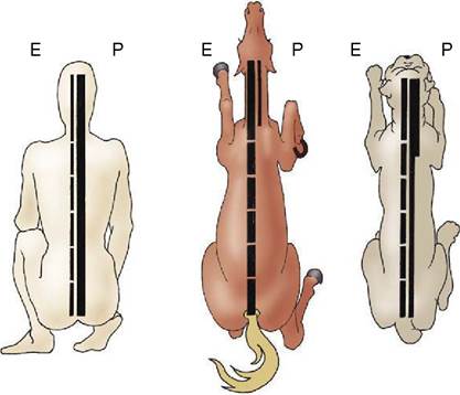
Figure 8-50 Comparison of the pyramidal (P) and extrapyramidal (E) systems of human, horse, and dog. The multisyn- aptic composition of the extrapyramidal system is indicated by the interruptions in this column; the width of the columns is an indication of their importance.
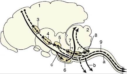
Figure 8-49 Relay diagram of the pyramidal (continuous line) and the extrapyramidal (interrupted line) systems. 1, Motor cortex; 2, cerebellum; 3, basal nuclei; 4, substantia nigra (mesencephalon); 5, red nucleus (mesencephalon); 6, pontine nuclei (metencephalon); 7, reticular formation; 8, olivary nucleus; 9, rubrospinal tract. a, Corticospinal fibers; b, corticobulbar fibers; c, corticopontine fibers.
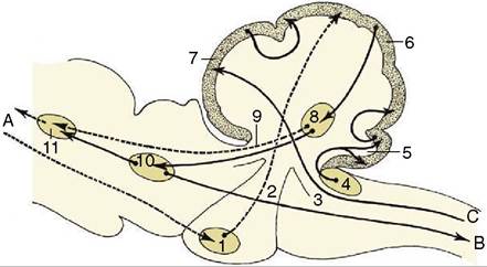
Figure 8-51 Some important fiber connections of the cerebellum. The connections with the neocortex are represented by broken lines. A, Tracts to and from the neocortex; B, tracts to the motor column of the spinal cord (extrapyramidal); C, proprioceptive tracts. 1, Pontine nuclei; 2, middle cerebellar peduncle; 3, caudal cerebellar peduncle; 4, cochlear nuclei; 5, flocculonodular lobe of the cerebellum; 6, neocerebellum; 7, rostral cerebellar lobe; 8, cerebellar nuclei; 9, rostral cerebellar peduncle; 10, red nucleus; 11, thalamic nuclei.
to their origin.
In domestic species, as in the generality of mammals, a short interneuron is always interposed; this interneuron is omitted from certain connections of the primate system.There are other differences among species. In primates and carnivores, pyramidal fibers reach all levels of the cord; in the dog about 50% terminate in cervical segments, 20% in thoracic segments, and 30% in lum- bosacrocaudal segments. In contrast, the pyramidal system of ungulates appears to have terminated by the level of origin of the brachial plexus (Figure 8-50), although there are hints of a diffuse continuation to lower levels within the dorsal funiculus—the route, incidentally, that is favored by the entire system in rodents. The proportion of fibers that decussate within the medulla oblongata also varies: about 50% do so in ungulates, 75% in primates, while all, or almost all, do so in the dog and cat.
The corticopontine fibers end within nuclei of the ventral pons; the axons of the neurons of the second order then decussate and pass within the transverse lamina of the pons to enter the cerebellum through its middle peduncle. Further successive synapses occur within the cerebellar cortex and then within the nuclei of the cerebellum, whence the return to the cerebral cortex is completed by relay through ventral thalamic nuclei (Figure 8-51). This arrangement constitutes the pyramidal feedback system.
The Extrapyramidal System
The extrapyramidal motor system includes all brain areas involved in regulating motor functions that are not included within the pyramidal system. It is more complicated and involves various multisynaptic pathways that relay within a series of nuclei dispersed through the brain from the telencephalon to the medulla oblongata. Some of these nuclei are large, grossly visible structures; others are small or diffuse, constituting a descending reticular system within the reticular formation of the brainstem. Tracts originating in the tectum and in the lateral vestibular nucleus are dealt with under visual and vestibular pathways (p.
299).The extrapyramidal system also takes origin from various parts of the cortex, including the primary motor area. The relay stations include the caudate nucleus among the basal nuclei, small subthalamic nuclei, the substantia nigra and red nucleus of the mesencephalon, the reticular formation, and the olive in the medulla oblongata (see Figure 8-49). Only the reticular formation and the red nucleus contain neurons that project directly (via interneurons) on the lower motor neurons of the brainstem and spinal cord; the other nuclei and also some neurons within the red nucleus project only on cells within nuclei lower in the series.
The fibers from the red nucleus decussate at once before descending through the ventrolateral part of the medulla oblongata to constitute a discrete (rubrospinal) tract bordering on the lateral corticospinal tract within the lateral funiculus of the cord (Figure 8-18Z√). This tract reaches to the most caudal part of the cord, projecting en route on ventral column cells (lower motor neurons) via short interneurons. This is an important tract in carnivores and is the best developed of all motor pathways in ungulates (see Figure 8-50). It serves as a modulator of pattern generators that are located in the spinal cord itself.
The reticulospinal system is divided between well- defined dorsal and ventral tracts located within the lateral funiculus and a third (pontine reticulospinal) tract within the ventral funiculus.
The activities of the various nuclei and connecting tracts of the extrapyramidal system are closely coordinated and so finely balanced that damage to any part may seriously impair the ability to maintain posture or to execute intended movements. Different parts of the system play different roles: some are facilitatory, others inhibitory, and yet others facilitatory through removal of other inhibitory influences.
The numerous feedback circuits associated with the extrapyramidal system maintain the necessary balance between these facilitatory and inhibitory influences.
The various circuits are, however, all subordinated to the overall control of the cerebellum to which all nuclei of the system project via relays within the olivary nuclear complex (see Figure 8-49). This complex projects, by way of the caudal cerebellar peduncle, to the contralateral cerebellar cortex before returning from the cerebellum to the various nuclei. The most important return limb runs from the cerebellum to the thalamic nuclei and thence to the motor cortex and basal nuclei; other pathways take shorter courses to project on the red nucleus and reticular formation.Cerebellar Function
Although the cerebellum does not itself initiate movement, it ensures that movements are executed as intended by controlling both pyramidal and extrapyramidal systems. To this end, it receives a continuous stream of information that flows from the pyramidal and extrapy- ramidal feedbacks to the caudal lobe, from the vestibular apparatus via the vestibular nuclei to the flocculonodular lobe, and from proprioceptors that feed into the rostral lobe.
The directions that are based on the integration of these various inputs within the cerebellar cortex are relayed through the cerebellar nuclei before issuing through the various peduncles to the contralateral red and thalamic nuclei, the reticular formation, and the vestibular nuclei (for the coordination of vestibular reflexes).