SPECIAL SOMATIC AFFERENT PATHWAYS
The Visual Pathways
Visual information is conveyed from the retina by the optic nerve. After entering the cranial cavity by the optic
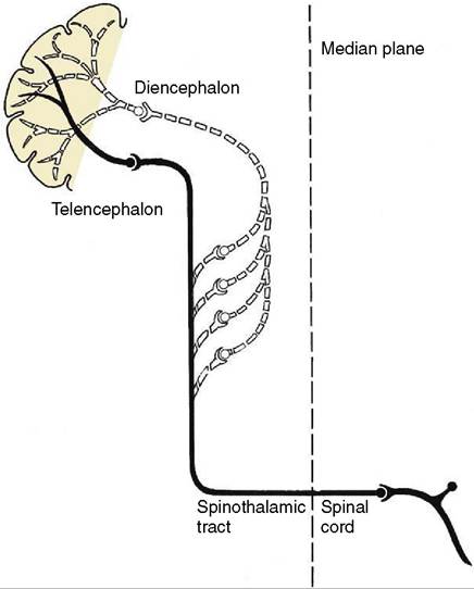
Figure 8-44 A multisynaptic ascending tract (white dotted line) to the telencephalon via the reticular formation.
The collateral tract in this example represents the extralemniscal projection (black).foramen, the nerve converges to meet its fellow in the optic chiasm on the ventral surface of the brain. Here, there is a partial decussation of fibers, and the proportion crossing has been correlated with the degree of binocular vision enjoyed by the species. In birds all fibers cross and vision was considered to be monocular; however, some recent information indicates that some birds have an even larger field of binocular vision than humans. In ungulates, the binocular field of vision is much restricted, and a very large percentage (85% to 90%) of fibers cross. A smaller proportion (75%) cross in carnivores, and about 50% cross in primates, in which binocular vision is best developed.
This reassortment brings fibers from both retinae into each optic tract, which arches over the lateral surface of the thalamus (Figure 8-23/20). The larger proportion terminates within the lateral geniculate nucleus, which raises a swelling on the upper end of the tract, or within the pulvinar nucleus medial to it. The primary optic pathway ends here. The fibers of the second-stage neurons project, via the optic radiation within the internal capsule, on the visual cortex, which is located within the occipital lobe of the cerebrum and is the seat of conscious visual perception (Figure 8-45/6).
A smaller number of the fibers project on various mesencephalic nuclei; some do so after preliminary relay in the lateral geniculate nucleus.
The foremost of these mesencephalic visual integration centers and nuclei are the rostral colliculi. From the mesencephalic nuclei there are relays through various neuronal chains by which the various visual and optic reflexes—concerned with direction of gaze, accommodation, and pupillary diameter—are effected. Fibers from the rostral colliculi also end on lower motor neurons in the cervical spinal cord and constitute the tectospinal tract, part of the so-called extrapyramidal system.Vestibular Pathways
The vestibular fibers enter the brainstem within the common vestibulocochlear trunk that penetrates the trapezoid body. They then terminate on, or detach collateral branches to, neurons of the vestibular nuclei (Figure 8-46/2). Those that continue unbroken reach the cerebellum by way of the caudal peduncle. The secondary fibers from the vestibular nuclei are divided between those that also pass to the cerebellum and the remainder, which run to the spinal cord via the vestibulospinal tract and medial longitudinal fasciculus. Within the cord they project via a series of interneurons on lower motor neurons of the ventral column. Other fibers proceed to the nuclei of the cranial nerves supplying the external ocular muscles; they follow the medial longitudinal fasciculus (Figure 8-46/4) and the reticular
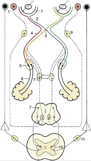
Figure 8-45 A simplified schema of the visual and pupillary reflex pathways. Thick lines, special somatic visual fibers; thin lines, sympathetic fibers; broken lines, parasympathetic fibers. 1, Retina; 1', dilated and constricted pupils; 2, optic nerve; 3, optic chiasm; 4, optic tract; 5, lateral geniculate nucleus; 6, optic radiation; 7, rostral colliculus and pretectal nuclei; 8, oculomotor nucleus (parasympathetic part); 9, ciliary ganglion; 10, lateral visceral efferent column; 11, cranial cervical ganglion.
formation. These tracts are part of the extrapyramidal system.
The fibers that lead to conscious perception of vestibular stimuli proceed via the lateral lemniscus and thalamic nuclei to a particular region of the cerebral cortex of the temporal lobe.
Auditory Pathways
The fibers of the cochlear component of the vestibulocochlear nerve relay within the dorsal and ventral cochlear nuclei located on the surface of the brainstem (Figure 8-47/1,2). The second-stage fibers from the ventral nucleus then proceed to a further synapse within an ipsilateral or contralateral nucleus of the trapezoid body (Figure 8-47/5). The pathway is then continued by fibers of third-stage neurons carried within the lateral
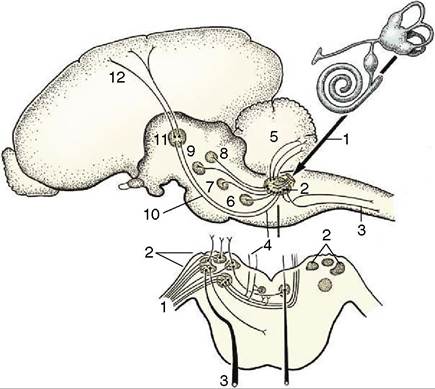
Figure 8-46 A simplified scheme of the vestibular pathways. 1, Vestibular fibers in vestibulocochlear nerve; 2, vestibular nuclei; 3, vestibulospinal tract; 4, medial longitudinal fasciculus; 5, vestibulocerebellar tract; 6, abducent nucleus; 7, trochlear nucleus; 8, oculomotor nucleus; 9, red nucleus; 10, vestibulothalamic tract (in lateral lemniscus); 11, thalamic nuclei; 12, thalamocortical projection fibers.
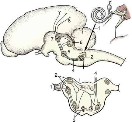
Figure 8-47 A simplified scheme of the auditory pathways. 1, Cochlear fibers in the vestibulocochlear nerve; 2, cochlear nuclei (dorsal and ventral); 3, nuclei in trapezoid body; 4, lateral lemniscus; 5, nucleus in lateral lemniscus; 6, caudal colliculus; 7, medial geniculate nucleus; 8, projection fibers for conscious perception.
lemniscus. A proportion of these synapse within the nucleus of this tract, a second contingent proceeds to the caudal colliculus (Figure 8-47/6), and a third, concerned with the conscious perception of sound, synapses in the medial geniculate nucleus before going to the auditory cortex, which is located within the temporal lobe.
The fibers that emerge from the dorsal cochlear nuclei join the ipsilateral or contralateral lateral lemniscus and thereafter follow the same courses as those that proceed from the ventral cochlear nuclei.