Some recapitulation and amplification of the earlier account (p. 7) of basic skin structure is now required.
Skin is composed of two parts: a superficial epithelium (epidermis) and a tough fibrous layer (dermis) that rests on a stratum of loose connective tissue (subcutis) (see Fig. 1.7).
The epidermis is continuously renewed. The surface cells are sloughed in flakes (e.g., dandruff) or as smaller particles (those of human skin accounting for much household dust) and are replaced by cell division in the deepest layer followed by migration of daughter cells toward the surface. As the epidermal cells migrate superficially, they undergo a series of molecular changes leading to their deaths, thus rendering them incapable of reacting to the various influences once they reach the surface. The sequence of changes, shown in Fig. 10.1, imposes an obvious stratification. The deepest layer (stratum basale) is closely molded on the irregularities of the underlying dermis and has a considerably greater area than the surface of the body (Fig. 10.1/1). As the cells move into the stratum spinosum, they shrink and draw apart, though remaining connected by intercellular bridges (desmosomes). The process of keratinization (cornification) now begins, and in the next layer (stratum granulosum) the cells contain scattered keratohyalin granules (Fig. 10.1/4). In some regions this layer is followed by a narrow stratum lucidum composed of flattened cells that have already lost their nuclei and distinct outlines but obtain a homogeneous appearance from the even dispersal of the granules. Finally, the outermost squamous layer (stratum corneum; Fig. 10.1/6) is densely packed with the fibrous protein keratin, the true horny substance, which is transformed keratohyalin. It is keratin that gives epidermal specializations (e.g., hair, hoof, and horn) their hardness and their strength.
The epidermal layers are thickest and most clearly differentiated where the skin is exposed to hard usage, as on the footpads of a dog (Fig.
10.2). Where abrasion is less severe, as in haired regions, the epidermis is much thinner, and neither the stratum granulosum nor the stratum lucidum may be clearly represented. The thickness of the epidermis depends on the mitotic rate within the stratum basale, which is adjusted by a substance (epidermal chalone) that inhibits cell division. Although cell production and loss normally match to maintain an even epidermal thickness, this balance may be disturbed in certain circumstances.There are no blood or lymphatic vessels in the epidermis, which is nourished by diffusion from the subjacent dermis.
The dermis is largely composed of collagen bundles, thickly felted together, as can be demonstrated by teasing leather (tanned dermis). Elastic fibers, which are also present, make the skin pliable and are able to restore its shape after being wrinkled or deformed. These fibers also draw apart the edges of a wound, making it gape (Fig. 10.3). Chronic tension damages the structure of the dermis, rupturing the connective tissue bundles; subsequent repair is usually by lighter scar tissue. The white lines (striae) of abdominal skin that appear after the completion of a pregnancy, especially in women, is an example.
The dermis is generously vascularized and innervated. It is also invaded by hair follicles and sweat, sebaceous, and other glands growing from the epidermis (see Fig. 1.7).
The interface for diffusion between the epidermis and the dermis is enlarged by the complicated molding of these components. The finger-like and ridgelike projections (papillae; Fig. 10.1/1') of the dermis fit closely into reciprocal depressions of the epidermis, and under normal conditions adhesion between the two structures is not easily disturbed. Trauma, such as that caused by the rubbing of an ill-fitting boot or shoe, sometimes separates them forcibly, and interstitial fluid then collects in a blister. The raw surface of the dermis exposed following the rupture of the blister normally is rapidly covered by the growth of the epithelium from the margins.
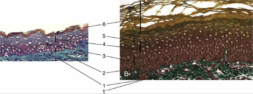
FIG.
10.1 Structure of the adult skin (Crossmon). (A) Skin from the canine flank. (B) Skin from a worn feline footpad; note the increased keratinization and the presence of a stratum lucidum and dermal papillae. 1, Dermis; 1', dermal papilla; 2, stratum basale; 3, stratum spinosum; 4, stratum granulosum; 5, stratum lucidum; 6, stratum corneum.FIG. 10.2
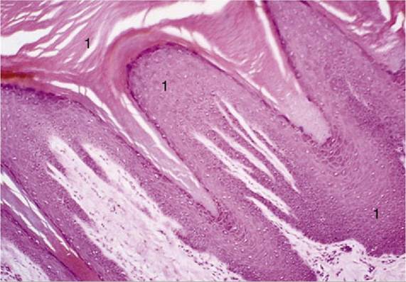
Stratified squamous epithelium of a footpad of a dog (hematoxylin and eosin; magnification
?70). 1, Very thick stratum corneum.
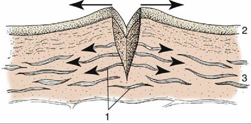
Skin incision; elastic fibers in the dermis cause the wound to gape (arrows). 1, Elastic fibers; 2, epidermis; 3, dermis.
FIG. 10.3
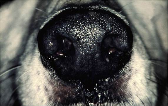
FIG. 10.4 The nose print in the dog can be used for identification of an individual.
The larger dermal ridges and papillae, generally developed where the covering epithelium is thickest, are reflected by corresponding epidermal contours. These contours are permanent and individually distinct and provide a means of identification, widely used in ourselves (fingerprinting) and less commonly used in other species (nose printing of dogs and cattle; Fig. 10.4).
The subcutis consists of loose connective tissue interspersed with fat. It varies in amount according to situation and is thin or even absent where movement is undesirable (e.g., over the lips, eyelids, and teats). It is particularly ample in dogs and cats, whose easily shifted skin can be grasped in large folds over much of the body (Fig. 10.5). In the pig and the human, the subcutis contains more substantial accumulations of fat, even in relatively ill-nourished individuals; this part constitutes the panniculus adiposus familiar in sliced bacon.
The clinical significance of the effects of dehydration or edema of the subcutis has been mentioned (p.
7).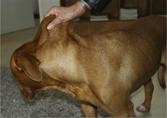
FIG. 10.5 Loose skin on the neck of a dog. Ample subcutis permits shifting of the skin.
The cutaneous blood vessels come from the vessels that supply the fasciae and superficial muscles. The arteries form a series of networks within the dermis. The most superficial network lies at the bases of the papillae and provides end-arteries that enter the papillae to release numerous capillaries that nourish the basal epidermal cells. Other capillary plexuses surround the hair follicles and associated glands (see Fig. 1.7). When the body temperature is raised, vasodilation of superficial vessels promotes heat loss—directly by surface radiation and indirectly by favoring the activity of the glands that produce sweat, which then evaporates. Conversely, the surface vessels constrict in cold environments or when the internal temperature drops. The blood flow is partly regulated by opening or closing of numerous anastomoses connecting the cutaneous arteries with veins. The skin vessels normally contain a considerable volume of blood, but much can be recalled to the musculature and internal organs after hemorrhage or shock.
Skin has a rich sensory innervation. The nerves accompany the vessels through the fasciae and form networks within the dermis. From these, fibers disperse to a variety of sensory receptors; some even penetrate a little way into the epidermis (see Fig. 9.33). Other (autonomic) fibers regulate the caliber of the smaller vessels, control the activity of skin glands, and excite the arrector pili muscles that attach to the hair follicles.
The epidermis develops from the embryonic ectoderm. This structure is initially a single layer of cells lying on a bed of mesenchyme that in time gives rise to the dermis (Fig. 10.6A). Long before birth the ectodermal cells begin to proliferate, pushing new cells toward the surface to produce a multilayered epithelium, while local condensations grow into the mesenchyme as the epithelial buds from which hair and glands differentiate. By the time of birth the skin of domestic mammals has a basically adult character, unlike that of many rodents and other small mammals that are born naked.
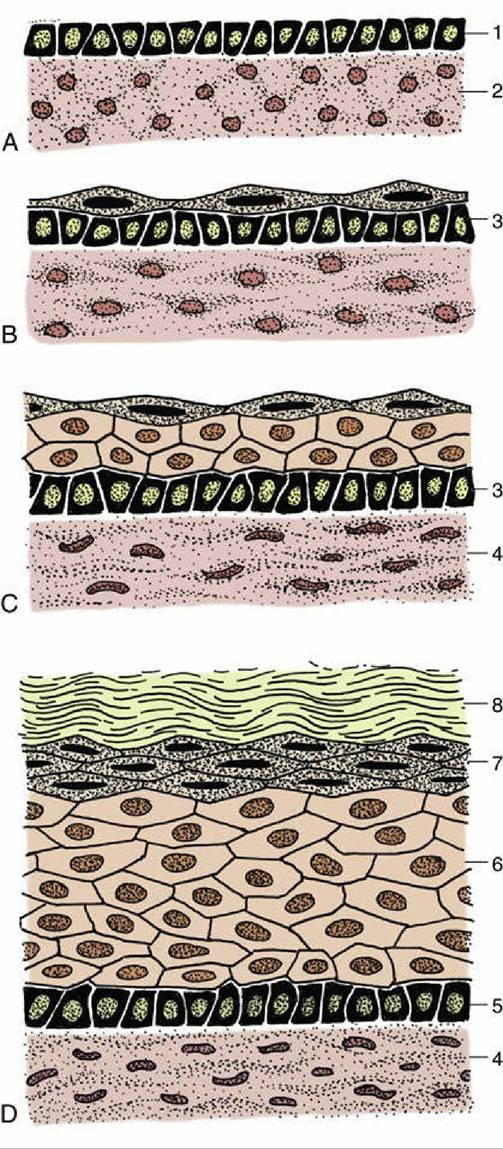
FIG. 10.6 Development of skin, schematic. (A) Skin of an early embryo. (B) Differentiation of epidermis and dermis. (C) Further differentiation of the epidermis. (D) Complete differentiation of the epidermis and dermis. 1, Ectoderm; 2, mesoderm (mesenchyme); 3, primitive stratum basale; 4, dermis; 5, stratum basale; 6, stratum spinosum; 7, stratum granulosum; 8, stratum corneum.