Hair is a mammalian feature, diagnostic of the class.
In most species a thick haircoat is spread over the body, except about the mouth and other openings and on the surfaces of the feet; in a few, including the domestic pig (though not its ancestors), the covering is sparse (see Fig.
10.10E). The individual hairs take a variety of intergrading forms, but only three need be distinguished here: straight, rather stiff guard hairs provide a "topcoat"; fine, wavy wool hairs provide an "undercoat"; and stout tactile hairs of restricted distribution are associated with touch receptors.Guard hairs mostly lie close against the skin and sweep uniformly in broad tracts, giving the coat a smooth appearance disturbed only by the whorls, crests, and partings formed where different streams converge and combine or diverge from one another. The regularity of the arrangement is significant because it promotes the runoff of rain, preventing the chilling that would occur if water were allowed to penetrate the pile to reach the skin. Occasionally, animals are born with a disturbed coat pattern, which may seriously impair their ability to withstand severe weather.
Each hair grows from a tiny pit or follicle to protrude above the surface of the skin. The follicle develops from an ectodermal bud that grows into the underlying mesenchyme during the embryonic stage of life. The bud branches give rise to skin glands (Fig. 10.7). The distal end of the bud forms a bulbous enlargement, which is then indented by a mesenchymal (dermal) papilla to form a primitive hair follicle. The epithelial cells lying against the papilla multiply, forming a hair matrix; the cells produced here keratinize and combine to form a primitive hair that grows through the center of the bud until it rises above the epidermis on the surface of the skin. Its passage takes it past the sebaceous glands that develop to the side of the follicle, and this arrangement allows the hair to obtain the oily coating so important for its health.
Although the ectoderm differentiates in this way, the mesoderm also condenses so that the tiny sheath around the embedded part of the hair acquires an outer mesodermal component.Fig. 10.8 shows only the essential features of a hair. It must suffice here to say that, in essence, a hair consists of a flexible column of closely consolidated and heavily keratinized, and hence dead, epithelial cells. Their arrangement permits the distinction of a medulla or core, a cortex, and an outer "scaly" cuticle. The variation in proportions and the arrangement of the parts permit the microscopic determination of the origin of a hair sample. In general, hairs with a thick medulla are straight and rather brittle, whereas those in which the cortex predominates are stronger and more pliable.
The proximal end of the follicle is joined by a tiny arrector pili muscle passing from an attachment near the dermal papillae (Fig. 10.7/8). The involuntary contraction of this muscle, which may be stimulated by a low ambient temperature, causes erection of the hair; the en masse erection of hairs traps more air and improves the insulation of the body. Although functionally unimportant in the human species, the effect is very obvious in our relatively naked skin when little mounds (goose pimples) appear over the courses of the arrector muscles. The fight-or-flight reaction mediated by the sympathetic nervous system raises the hackles that give an animal a threatening appearance.
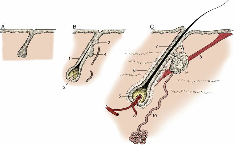
FIG. 10.7 Development of hair and associated sebaceous and sweat glands, schematic. (A) Ectodermal bud growing into mesenchyme. (B) Differentiation of the bud; indications of glands appear. (C) Hair follicle with accessory structures. 1, Primitive hair follicle; 2, dermal papilla; 3, bud of sweat gland; 4, bud of sebaceous gland; 5, bulb (hair matrix) of hair; 6, hair follicle; 7, root of hair; 8, arrector pili muscle; 9, sebaceous gland; 10, sweat gland.
In the adult, many glands open independently, not into hair follicles.There are many local variations in the form and development of guard hairs. Familiar examples are the stiff, sparsely scattered bristles of pigs (see Fig. 10.10E), the coarse hair of the mane and tail of horses, the long tail hairs of cattle, the fetlock tufts of horses, and the feathering of the tail and limbs of certain breeds of dogs. The hormone-dependent local variations particularly evident in the human species include the male beard and the sexually dimorphic distribution of body hair. Baldness as an accompaniment of advancing age is especially a problem of the human male. Testosterone, which is responsible for the growth of the beard and coarse body hair, paradoxically seems to trigger early baldness in genetically predisposed individuals; a reduced blood level of thyroxine, which initiates and controls hair growth, also plays some part.
Hairs have restricted lives and are discarded sooner or later. Although hair shedding in humans is a continuous process involving only a few hairs at a time, most other species, especially wild species, shed many hairs at a time in a seasonal fashion. Even domesticated animals protected from the more extreme climatic changes show a recurrent pattern with peaks in the spring and fall; spring shedding lasts about 5 weeks in dogs. Shedding is more obvious in animals not regularly groomed to remove dead hair. Cats also molt most heavily in spring, with less substantial loss through the summer and fall followed by attaining of prime condition in winter. For the same reason, the pelts of furbearing species are harvested in winter, although the number of harvested pelts has fallen with changing social attitudes.
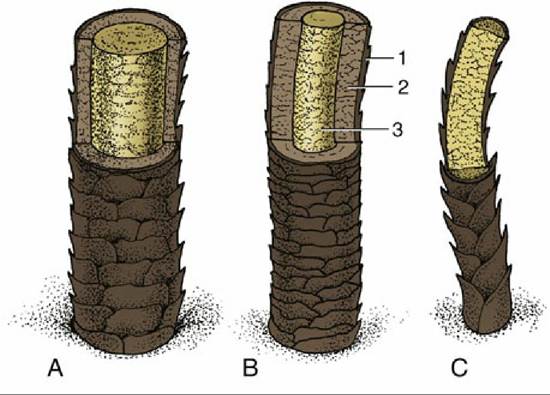
FIG. 10.8 Schematic representation of three kinds of hair. (A) Guard hair with thick medulla. (B) Guard hair with thick cortex and thin medulla.
(C) Wool hair; the cortex is absent. 1, Cuticle; 2, cortex; 3, medulla.The seasonal replacement begins with a slowing of the growth of existing hair mainly owing to a rise in seasonal temperature. As growth slows (in the so-called catagen phase) the hair matrix and covering papilla both atrophy (Fig. 10.9B). No growth occurs in the ensuing (telogen) phase when the follicle, including the papilla, shortens, causing a larger part of the hair to project above the skin in simulation of growth (Fig. 10.9D). When growth resumes, the follicle, with its matrix now reactivated, lengthens, and as it again extends away from the surface, it loses its grip on the old hair, which falls out. A replacement hair then forms in the active growth (anagen) phase that follows to emerge on the surface of the skin.
Wool hairs provide the soft undercoat. They are thin, wavy, and in most species, shorter and more numerous than the guard hairs by which they are concealed. The distinction between hair fiber types is not always clear-cut, and intermediate forms exist to complicate description, especially in the sheep.* Wool is not, of course, confined to sheep among domestic animals. Cashmere and Angora goats, Angora rabbits, and alpacas all produce wools of distinctive quality that are utilized in the production of luxury yarns and textiles.
In many species, including mature dogs and cats, several hairs share a single follicle opening (Fig. 10.10B-D). The central (primary) hair is longest and of the guard type, but the surrounding (secondary) hairs are shorter and softer; they provide the undercoat and may be designated wool hairs because they have little medulla.
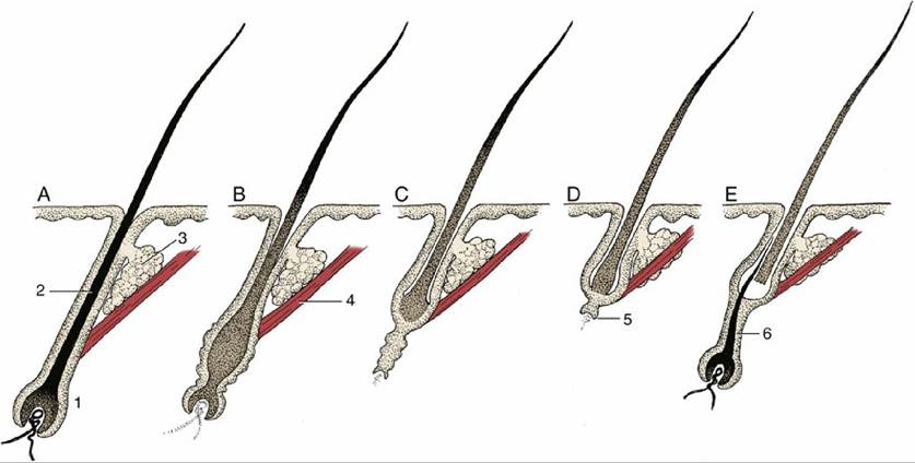
FIG. 10.9 Phases of the hair cycle. (A) Fully functional hair follicle; anagen phase. (B) Follicle begins to atrophy; early catagen phase. (C) Further atrophy of follicle; late catagen phase. (D) Atrophied follicle; hair is displaced distally and new hair matrix begins to form; telogen phase.
(E) New hair matrix established and new hair begins to grow; early anagen phase. 1, Hair follicle; 2, root of hair; 3, sebaceous gland; 4, arrector pili muscle; 5, new hair matrix; 6, new hair.Tactile hairs are substantially thicker and generally protrude beyond the neighboring guard hairs. Most are found on the face, principally on the upper lip and about the eyes, although others are scattered (in species-variable fashion) on the lower lip, the chin, and elsewhere on the head. The cat, whose whiskers are particularly good examples (Fig. 10.11), also possesses a cluster of similar hairs at the carpus. Tactile hair follicles reach deep into the subcutis or even the superficial muscles. They are characterized by the presence of a venous sinus filled with blood and located between inner and outer layers of the dermal sheath (Fig. 10.12). The nerve endings responsive to mechanical stimulation are also contained within the dermal sheath (Fig. 10.12A). The stimulus provided by disturbance of the hair is amplified by wave motion in the blood. The follicles of tactile hairs appear early in development, before those of the coat hairs, and their staged appearances provide useful criteria for aging embryos.
The skin of dogs and cats presents minute scattered tactile elevations (toruli tactiles) usually associated with special (tylotrich) guard hairs; the roots of these are surrounded by venous sinuses similar to, though smaller than, those of true tactile hairs. These elevations are also sensitive to touch (Fig. 10.13).
The restricted distribution of various pigments creates a distinct pattern of coats as seen in breeds such as Holstein cattle and Dalmatian dogs. The pigments, polymers of melanin ranging from black, through brown and red, to lighter shades are present in granule form* within cells of the epidermis, hair follicles, and hair. The pigments protect the skin from potentially harmful ultraviolet radiation and are unnecessary within those epidermal regions that are covered by a dense coat of hair.
In most mammals, unlike in humankind, skin pigmentation is therefore restricted to a few exposed parts that include the modified area associated with the external nose. It may be lacking here in white-coated individuals that obtain equivalent protection from a thickened stratum corneum.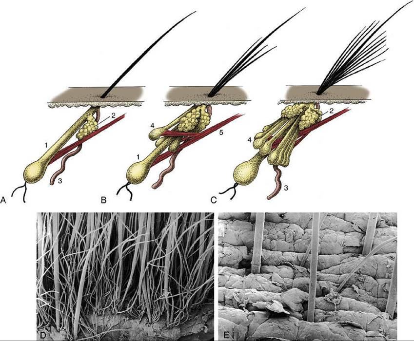
:IG. 10.10 Hair follicles of the dog: (A) simple follicle present shortly after birth; (B) follicle present durin< the first few months after birth; (C) complex adult follicle; the primary hair is surrounded by several secondary hairs. (D) Scanning electron micrograph of adult canine skin; note one or two follicles without primary (guard) hairs. (E) “Naked” skin of a pig with sparse primary hairs (bristles) and surface debris. 1, Primary hair follicle; 2, sebaceous gland; 3, duct of sweat gland; 4, secondary hair follicle; 5, arrector pili muscle.
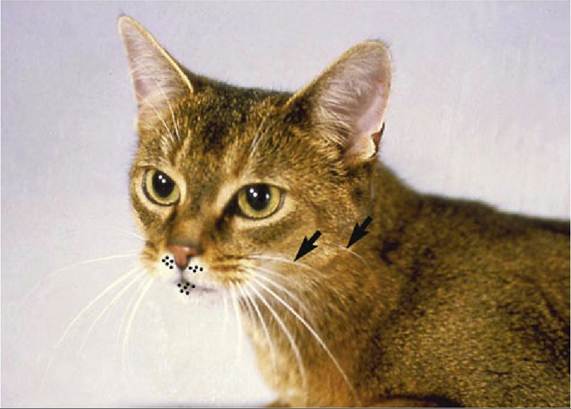
FIG. 10.11 Tactile hairs on the head of the cat. The dots on the lips show the position of the circumoral glands. The arrows point to the buccal (tactile) hairs.