Stimulation of Sensory Pathways Can Be Recorded as Evoked Potentials
Large areas of the brain and the spinal cord are not reflected in the EEG. Other clinical electrophysiological recordings can help examine the function of these areas.
Synaptic activity in a sensory pathway can be recorded from the scalp by a computerized technique that averages out the more random background EEG activity and averages in the electrical response to multiple stimulations of a sensory system.
Such signals are called sensory-evoked potentials.Because scalp macroelectrodes can more easily record the EEG electrical signals generated from the closer cerebral cortical cells, these higher-voltage signals must be eliminated; otherwise, they would mask the sensory-evoked potentials. Because the background EEG signals are relatively random, a
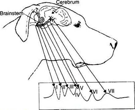
FIGURE 16-5 Brainstem auditory-evoked response (BSAER): idealized diagram of waveforms recorded by signal averaging. Neural elements believed to generate sequentially the auditory waves are grouped as follows: wave I reflects the cochlea, spiral ganglia, and cranial nerve VIII; wave Il reflects the cochlear nuclei; wave III reflects the nucleus of the trapezoid body; waves IV and V reflect the lateral lemniscus and lemniscal nuclei and caudal colliculus, respectively (these two waves are frequently combined to form one wave); wave Vl reflects the medial geniculate body; and wave Vll reflects auditory radiations. Positive is upward. (From Oliver JE, Hoerlein BF, Mayhew IG, editors: Veterinary neurology, Philadelphia, 1987, Saunders.)
computer can average them together and functionally erase them from the recording while at the same time averaging the nonrandom sensory-evoked potential signals recorded from multiple stimulations of a sensory pathway. In this way, scalp macroelectrodes can be used to record electrical events generated in brain locations distant from the recording electrode.
For this reason, these sensory-evoked potentials are often called far-field potentials.One such sensory-evoked potential is the brainstem auditory-evoked response (BSAER). This clinical electrophysiological procedure, in which the electrode placement is configured to include activity from the brainstem, records the electrical events for 10 msec after a click stimulus to the ear (Figure 16-5). Usually, seven waves are recorded, thought to be generated by neural activity in components of the auditory pathway from the auditory nerve through the auditory radiations leaving the medial geniculate nucleus of the thalamus. Recordings longer than 10 msec are sometimes taken. These later waves reflect cortical response to auditory stimulation. BSAER is used in animals and humans to assess brainstem function in general and auditory function in particular. Other sensory-evoked potentials can be recorded from the visual system, the somatosensory system, and other sensory modalities.
BSAER is also often used with simultaneous EEG recording in the confirmation of brain death. A flat EEG, a very crude indicator of brain death, combined with a viable BSAER can suggest that the functional deficit may not be irreversible.