The Collective Behavior of Cortical Neurons Can Be Studied Noninvasively Through the Use of Macroelectrodes on the Scalp
The scalp EEG records a fluctuating voltage resulting from changes in postsynaptic potentials in thousands of neurons below the electrode. Each change in voltage has a polarity.
By convention, changes in voltage measured by extracellular electrodes, such as those on the scalp, have a standard direction of recorder deflection.
When the voltage change is in a positive direction, the recorded deflection is “down”; when in a negative direction, the deflection is “up” (Figure. 16-4). The polarity of the voltage change at the scalp depends on the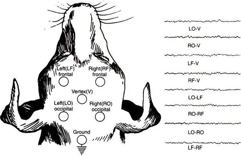
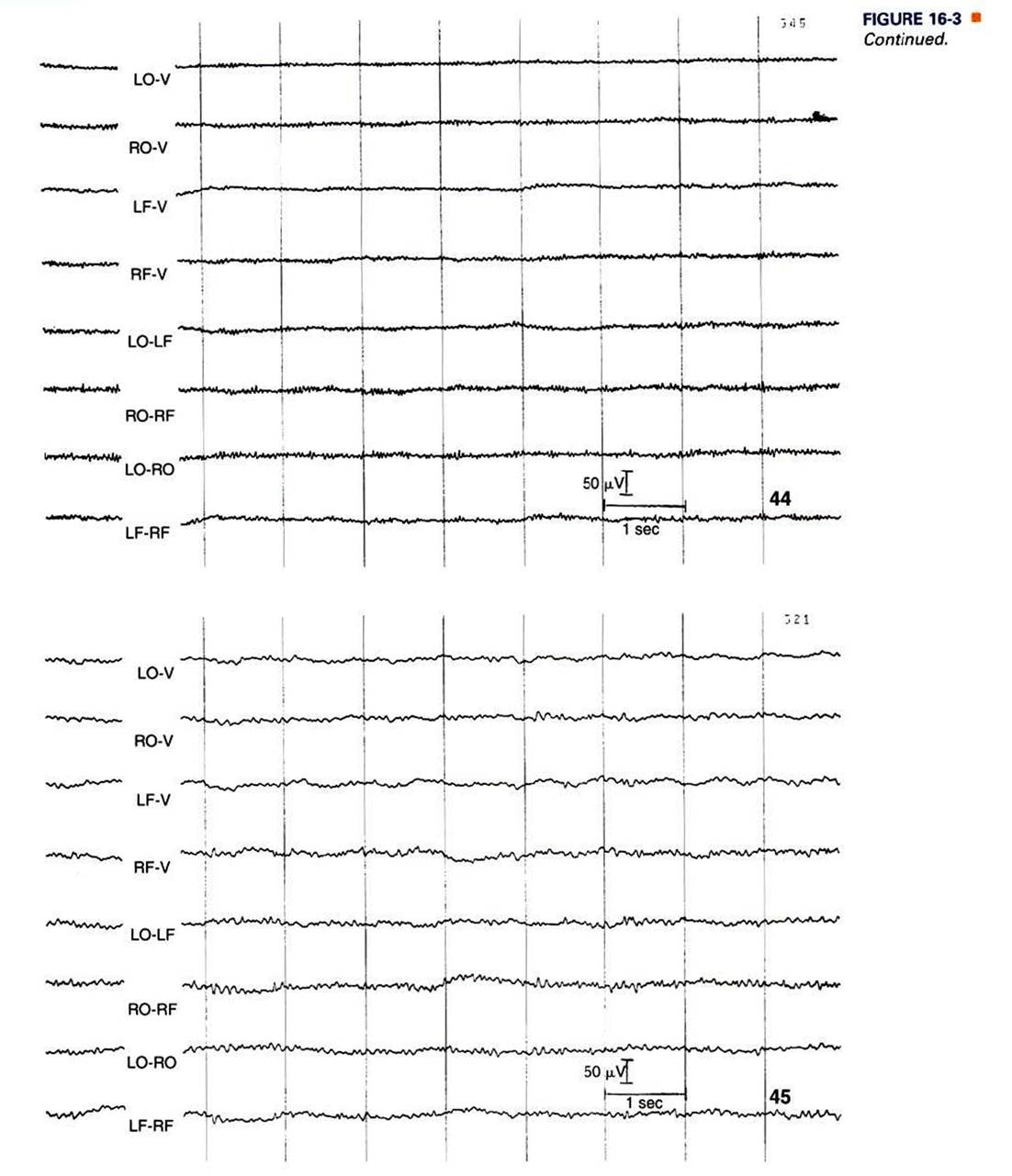
FIGURE 16-3 EEG recorded from various combinations of lead points using an older, simpler electrode configuration (the Redding configuration). It shows the difference in frequency and amplitude between an alert animal (44), a relaxed animal (45), and an animal in light sleep (80). Note the decrease in frequency and increase in amplitude in the progression from alert to relaxed to light sleep. (From Oliver JEr Hoerlein BF, Mayhew IG, editors: Veterinary neurology, Philadelphia, 1987, Saunders.)
Figure continues on next page
nature and location of the postsynaptic potential change. Ifan excitatory postsynaptic potential (EPSP) occurs in a deep cortical layer, positive ions (e.g., Na,) enter the cell there, leaving the extracellular fluid at that location relatively negative. Through principles of volume conduction, this leaves the extracellular fluid near the cortical surface positive with respect to the deeper, negatively charged region of extracellular fluid (see Figure 16-4; for simplicity, only one cell is indicated). This results in a positive voltage change being recorded at the scalp macroelectrode near the cortical surface.
Based on theThe Electroencephalogram and Sensory-Evoked Potentials CHAPTER 16
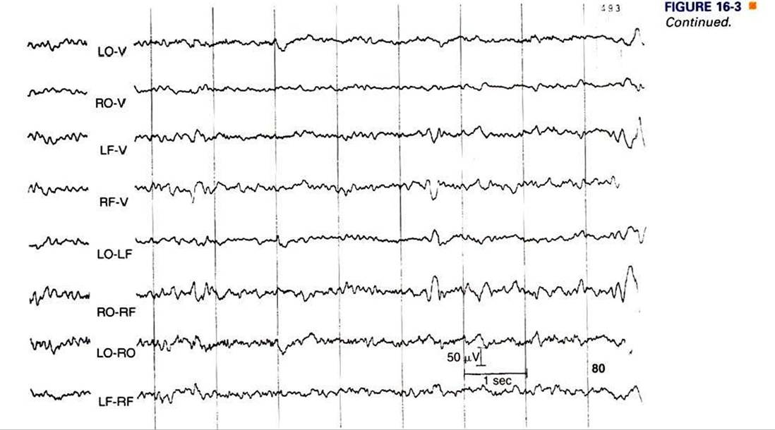
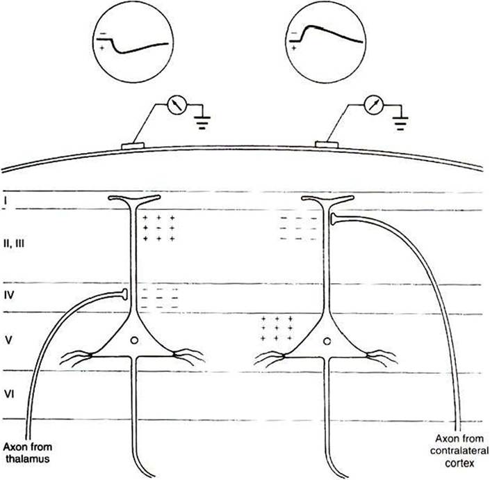
FIGURE 16-4 Scalp recordings and underlying synaptic mechanisms. Left, Potential recorded from a scalp electrode after activation of thalamic inputs.The terminals of thalamocortical neurons make excitatory connections on cortical neurons predominantly in layer IV.Thus the site of inward current flow (sink) in layer IV leaves the extracellular fluid at that location relatively negative and the extracellular fluid near the cortical surface relatively positive. Because the recording electrode is located on the scalp, near the cortical surface, it records a positive potential. By convention, a positive extracellularly recorded potential is, unlike intracellular recordings, a downward deflection. Right, Potential recorded from an excitatory callosal afferent originating in the contralateral cortex.The axon of this callosal neuron terminates in a superficial cortical layer. A negative potential (upward deflection) is recorded because the electrode is closer to the site of inward current flow, which leaves the extracellular fluid near the cortical surface relatively negative. (From Kandel ER, Schwartz JH: Principles of neural science, ed 2, NewYork, 1985, Elsevier Science Publishing.)
same principles, if the EPSP occurs near the cortical surface (see Figure 16-4), the voltage recorded from the scalp is negative. The polarity of these changes would be reversed for inhibitory postsynaptic potentials (IPSPs).
Voltage changes recorded from the scalp are the result of the summated extracellular voltage changes caused by the postsynaptic potentials of a large number of active cortical neurons, primarily pyramidal cells, because the voltage change from any one neuron is too small to record.
Action potentials contribute little to the EEG with scalp electrodes.The amplitude (height) of voltage fluctuations in the scalp- recorded EEG is a function of how many cortical cells are changing their postsynaptic potentials in the same direction at the same time. Because a high-amplitude voltage change would result from a large number of neurons firing synchronously, a high-amplitude, slow-frequency EEG is said to be a synchronized EEG. When neurons are firing more or less at random, a low-amplitude, high-frequency EEG results, said to be a desynchronized EEG.
The frequency with which EEG voltage changes occur is largely determined by the reticular activating system. As noted in Chapter 10, ascending projections of the reticular formation play an important role in modulating consciousness, arousal, and attention. Many of these projections synapse primarily in the thalamus, hypothalamus, or directly in the cerebral cortex in a diffuse fashion. Diffuse cortical projections from portions of the thalamus (intralaminar nuclei) and hypothalamus (lateral hypothalamus), along with diffuse, direct cortical projections from the reticular formation, likely regulate consciousness and arousal. Neurons that project to cortex from specific sensory relay nuclei of the thalamus and receive reticular formation input probably influence attention. The term reticular activating system collectively refers to these ascending reticular formation neurons and the neurons that relay their activity to the cortex, both of which affect consciousness, arousal, and attention.