SUPERFICIAL NERVES
Only a few features of the superficial nerves require notice. The facial nerve detaches its auriculopalpebral branch before it enters the face (see Figure 18-36∕2√). This branch then takes an independent course across the zygomatic arch (where it is palpable), which leads it between the eye and the ear.
The branch may be blocked by injection between the caudal end of the arch and the base of the ear. The procedure facilitates examination of the eye because it eliminates blinking and closure of the lids (p. 345).The facial trunk divides into dorsal and ventral buccal branches before or, more commonly, shortly after emerging from under the protection of the parotid gland (Figure 18-7/9). These branches and the smaller divisions into which they soon assort run forward over the masseter, where they are palpable and sometimes even visible through the skin. Blows over the masseter or pressure in prolonged recumbency may damage some or all of the divisions. The asymmetry of the face that results when the muscles of the lips, cheek, and nose are paralyzed is usually more striking than in other species. Because the auriculopalpebral branch is precociously detached, such trauma generally spares the muscles of the eyelids and external ear; their involvement points to injury at a more proximal level, which suggests a more sinister causation (Figure 18-8).
The sensory innervation of the face is the duty of the trigeminal nerve. It is an easy matter to locate some of
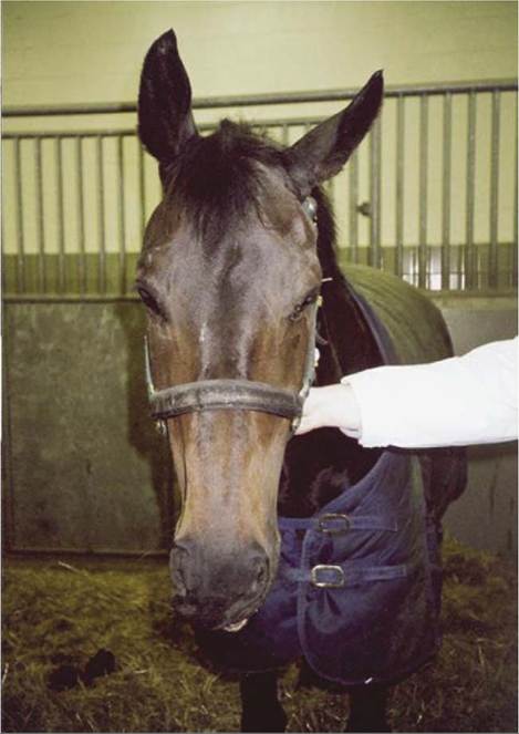
Figure 18-8 Injury to the facial nerve. Note pronounced drooping of ear, moderate drooping of upper eyelid of affected side, and distortion of the nose, which is drawn toward the sound side.
the principal branches concerned—the supraorbital, infraorbital, and mental nerves—where they emerge from the corresponding foramina (Figure 18-9/1,3).
The supraorbital nerve leaves the supraorbital foramen within an easily located dimple in the root of the zygomatic process of the frontal bone. The nerve supplies the upper eyelid and adjacent part of the forehead skin. Directions for location of the infraorbital and mental nerves have already been given (pp. 504 and 505). Anesthetic deposited about the infraorbital nerve at its emergence will desensitize the skin of the upper lip, nostril, and much of the nose extending well caudal to the foramen. Blockage of the mental nerve desensitizes the skin of the lower lip and chin region. During blockage of either of these nerves, it is possible to insert the tip of the needle through the foramen into the bony canal within the jaw. If this is done, injection of anesthetic will also deaden the more rostral teeth (from P2 forward).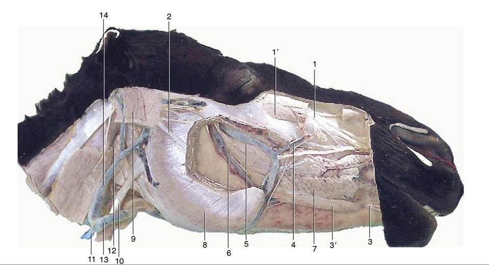
Figure 18-9 Deeper dissection of the head. Parts of the superficial muscles, masseter, and parotid gland have been removed.
I, Infraorbital nerve; 1', levator labii superioris; 2, dorsal buccal branch of facial nerve; 3, mental nerve; 3', depressor labii inferioris; 4, facial vein; 5, deep facial vein; 6, buccal vein; 7, buccinator; 8, masseter; 9, occipitomandibularis; 10, sternocephalicus;
II, external jugular vein; 12, mandibular gland; 13, linguofacial vein; 14, maxillary vein.