THE NASAL CAVITY
Some features of the external nose have been described (p. 501). The ventral part of the nostril leads through a constricted vestibule into a nasal cavity considerably less roomy than might be supposed from the exterior.
The factors that determine this are common to all species, but their importance is exaggerated in the horse by the reserve portions of the cheek teeth and the extensive development of the paranasal sinus system (see Figure 3-14).The dorsal and ventral conchae form delicate scrolls that coil in opposite directions from their lateral attachments (Figure 18-10). The space enclosed within each is divided into two compartments by an internal septum. The caudal part of the dorsal concha is occupied by a rostral extension of the frontal sinus with which it enjoys free communication. The caudal space within the ventral concha communicates with the rostral maxillary sinus. The space within the rostral part of each major concha is in direct communication with the nasal cavity. Numerous small ethmoidal conchae projecting into the fundus serve to enlarge the olfactory area (Figure 18-11/3).
The major conchae divide the cavity into the usual pattern of meatuses (see Figure 18-10). It may be presumed (for direct evidence is lacking) that air moves from the dorsal meatus to the olfactory mucosa and from the middle meatus to the sinuses, while the ventral and common meatuses supply the principal respiratory passage. The conjunction of the last two provides the widest and most convenient route for the introduction of a stomach tube, endoscope, or other instrument. The fragility of the ventral concha and the vascularity of the covering mucosa require that the procedure be performed with care.
Because breathing through the mouth is impossible, augmentation of the air intake in conditions of stress depends on reduction of the obstruction offered by the nose itself. The nostrils may be greatly widened by obliteration of the nasal diverticulum (see Figure 18-2), while contraction of the mucosal venous plexuses thins (and blanches) the membrane. Conversely, congestion of the mucosal vessels seriously impedes air flow. In infections, thickening of the mucosa around the slitlike entrance to the sinus system may obstruct its drainage, damming back a catarrhal exudate.
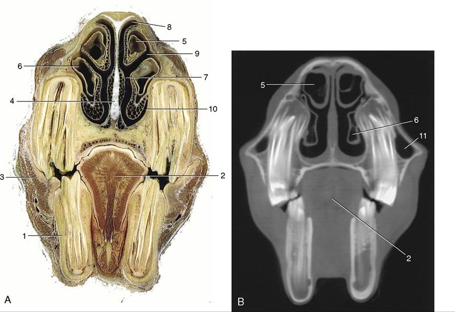
Figure 18-10 A, Transverse section of the head at the level of the rostral maxillary sinus. B, Computed tomographic scan (bone window) at about the same level. 1, P4; 2, tongue; 3, buccinator; 4, nasal septum; 5, dorsal nasal concha; 6, ventral nasal concha; 7, common nasal meatus; 8, dorsal nasal meatus; 9, middle nasal meatus; 10, ventral nasal meatus; 11, rostral maxillary sinus.
The vomeronasal organ does not communicate with the mouth in the horse but maintains the usual connection with the nasal cavity (Figure 18-12/2).