Superficial Vessels
The facial artery and vein enter the face in company with the parotid duct (Fig. 18.7/8). The artery is easily found and is convenient for taking the pulse (see Fig. 18.40/7), especially just before it crosses the lower border of the mandible (on the medial side of the mandible).
The artery then ascends along the rostral margin of the masseter before terminating in divergent branches with varying pattern. However, it is usually possible to identify the inferior and superior labial, lateral and dorsal nasal, and angularis oculi arteries.The arrangement of the veins is similar, and their pattern may be visible in life in thin-skinned horses. Certain of the tributaries turn caudally, deep to the masseter, to anastomose with other veins of the head. The most dorsal connection, the transverse facial vein (Fig. 18.8/4), joins the superficial temporal vein. The rostral part lies deep to the masseter while the caudal part lies superficially and follows the ventral edge of the zygomatic arch. The caudal stretch is accompanied by an artery (an alternative site for examination of the pulse) and a nerve. Another site for pulse taking is the subcutaneous segment of the masseteric artery (Fig. 18.7/12).
The second connection, the deep facial vein (Fig. 18.8/5), burrows below the masseter and perforates the periorbita before passing through the orbital fissure to join the cavernous venous sinus within the cranial cavity. Two features of this vein are believed to possess functional significance. The cavernous sinus contains relatively cool blood drained from the hard palate and nasal cavity. The sinus envelops the internal carotid artery and may cool the arterial blood flowing to the brain. Second, an expansion of the vein deep to the masseter may form the basis of a pumping mechanism. It is liable to compression by the masseter, and it is asserted that this helps prevent stagnation of the venous return from the lowered head of the grazing animal.
There is a similar expansion on the third connection, the buccal vein (Fig. 18.8/6), which also runs deep to the masseter to join the superficial temporal tributary of the maxillary vein.
There are two superficial groups of lymph nodes. The parotid group under cover of the rostral part of the parotid gland is not usually palpable unless enlarged. The second group comprises numerous mandibular nodes arranged in a spindle within the intermandibular space. Together with their contralateral fellows, these nodes form a forward-pointing "V" shape that is always very distinctly palpable (see Fig. 18.39/2). The course of the lymph flow is dealt with later (pp. 520-521).
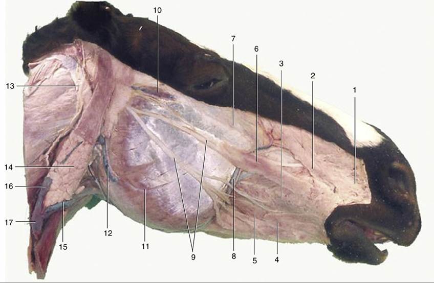
FIG. 18.7 Superficial dissection of the head. 1, Caninus; 2, levator nasolabialis; 3, buccinator; 4, stump of cutaneous muscle joining orbicularis oris; 5, depressor labii inferioris; 6, zygomaticus; 7, levator labii superioris; 8, facial artery and vein; 9, buccal branches of facial nerve; 10, transverse facial artery and vein and transverse facial branch of auriculotemporal nerve; 11, masseter; 12, masseteric artery and vein;
13, great auricular nerve (C2); 14, parotid gland; 15, linguofacial vein; 16, maxillary vein; 17, external jugular vein.