Sympathetic and Parasympathetic Nerves Act on Cardiac Pacemaker Cells to Increase or Decrease the Heart Rate
Figure 19-8 shows how the neurotransmitters norepinephrine and acetylcholine affect the pacemaker cells of the heart. Norepinephrine exerts its effect by activating β-adrenergic receptors on the cell membranes of pacemaker cells.
Activation of such receptors speeds up the ion channel changes that are responsible for the spontaneous depolarization of pacemaker cells. Because the pacemaker cells reach threshold more quickly in the presence of norepinephrine, there is a shorter interval between heartbeats. Therefore, heart rate is elevated above its intrinsic or spontaneous level.Acetylcholine has the opposite effect. Acetylcholine activates muscarinic cholinergic receptors on the cell membranes of pacemaker cells, which slows the ion channel changes that are responsible for the pacemaker cell’s spontaneous depolarization. Acetylcholine makes it take longer for pacemaker cells to reach threshold. There is consequently a longer time between heartbeats, so the heart rate is slowed below its intrinsic or spontaneous rate.
Sympathetic neurons release norepinephrine at the SA node cells, and thus sympathetic nerve activity increases the heart rate. Epinephrine or norepinephrine, released from the
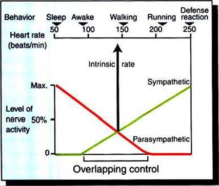
FIGURE 19-9 Upper scale shows that heart rate in a normal, large dog ranges from 50 to 250 beats/minute, depending on behavioral state.The graph shows that this wide range of heart rates is brought about by the interactions between sympathetic nerve activity, which speeds the heart above its intrinsic rate, and parasympathetic nerve activity, which slows the heart below its intrinsic rate. Sympathetic and parasympathetic nerves are simultaneously active over a considerable portion of the heart rate range (Overlapping control). Note that the heart beats at its intrinsic rate (about 140 beats/minute) either in the absence of any neural influence or when sympathetic and parasympathetic effects are equal and opposite.
adrenal glands and circulating in the bloodstream, has the same effect. Parasympathetic neurons release acetylcholine at the SA node cells, and thus parasympathetic activity decreases the heart rate. Figure 19-9 illustrates how sympathetic and parasympathetic neurons interact in the control of the heart rate. In the absence of norepinephrine and acetylcholine, the heart beats at its intrinsic rate. For a large dog, this rate is typically about 140 beats per minute (beats/min). Heart rates below the intrinsic rate are achieved by activation of parasympathetic neurons and release of acetylcholine. Accordingly, the graph indicates that parasympathetic activity is high during awake rest (heart rate of 90 beats/min) and very high during sleep (heart rate of 55 beats/min). Heart rates above the intrinsic rate occur during exercise or emotional arousal and are achieved by activation of the sympathetic nerves to the heart and release of norepinephrine (or by circulating epinephrine or norepinephrine). The highest possible level of sympathetic activity, and therefore the highest possible heart rate, occurs during maximal exercise or a defense alarm reaction (fear, “fight or flight” response).
Through variation in the levels of sympathetic and parasympathetic tone, the dog’s heart rate is adjusted, over a wide range, as appropriate for each behavioral situation. When both systems are partially active, the resulting heart rate represents the outcome of a “tug-of-war” between sympathetic action to increase the heart rate and parasympathetic action to decrease the heart rate. Typically, the sympathetic and parasympathetic systems are both partially active during awake
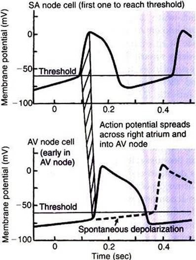
FIGURE 19-10 Both sinoatrial (SA) node cells and atrioventricular (AV) node cells exhibit pacemaker activity (spontaneous depolarization toward threshold). Normally the SA node cells depolarize more quickly and reach the threshold first (top).
The resulting atrial action potential propagates into the AV node (as represented by hatched band) and depolarizes the AV node cells quickly to their threshold, causing them to form an action potential (solid line, bottom graph). However, if the SA node pacemaker cells become nonfunctional or if atrial action potentials are not conducted into the AV node, the AV node cells eventually depolarize to threshold and initiate action potentials on their own (dashed line, bottom graph). In this way the AV node cells serve as an auxiliary (emergency) ventricular pacemaker.states, ranging from quiet rest (heart rate about 90 beats/min) to moderate exercise (heart rate about 175 beats/min). Parasympathetic activity predominates in the lower part of this range, and sympathetic activity predominates in the higher part. When sympathetic activity and parasympathetic activity are equal, their effects cancel, and the heart rate is at its intrinsic (spontaneous) level. Simultaneous activation of sympathetic and parasympathetic neurons appears to give the nervous system tight control over the heart rate under a wide variety of behavioral conditions.