Systemic Circulation
Systemic circulation refers to the movement of oxygenated blood to all areas of the body and the return of deoxygenated blood to the heart (Fig. 17-5). The following descriptions of the blood vessels of the systemic circulation are based mainly on the horse (Fig.
17-6). Animals having different digestive systems, such as ruminants, and those having more than one digit per limb of course have a somewhat different arrangement of arteries and veins in association with their specific anatomy.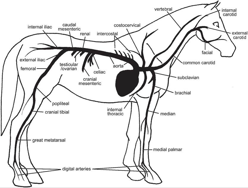
Figure 17-6. Main arteries of the horse.
Aorta
Aortic Arch. The left ventricle receives oxygenated blood from the left atrium and pumps the blood throughout the systemic circulation by way of the largest artery, the aorta. The aortic valve, at the junction of the left ventricle and aorta, prevents backflow of blood from the aorta into the left ventricle when the ventricle relaxes.
Two large vessels arise from the aorta immediately distal to the aortic valve. These are the right and left coronary arteries, and they are the arterial blood supply for the myocardium. Most of the venous blood from the myocardium is returned to the right atrium by way of the coronary veins, which empty directly into the right atrium by way of the coronary sinus, adjacent to the opening of the caudal vena cava.
After emerging from the base of the heart, the aortic arch courses dorsad and then caudad, just ventral to the bodies of the thoracic vertebrae. The aorta continues as the thoracic aorta until it passes through the aortic hiatus of the diaphragm to become the abdominal aorta. Arteries that supply the head, neck, and thoracic limbs branch from the aortic arch.
in horses and ruminants, the aortic arch gives rise to a single large brachiocephalic trunk, whose many branches distribute blood to the cranial half of the animal.
The precise pattern of the main arterial branches is species dependent, but the following generalities can be made: (1) the main blood supply to the thoracic limbs arises as right and left subclavian arteries; (2) the right and left costocervical trunks provide arterial blood to regions of the neck and cranial thoracic wall; and (3) right and left common carotid arteries, a main source of blood for the head and brain, arise together from a single bicarotid trunk.Thoracic Aorta. The thoracic aorta passes caudad just ventral to the vertebral bodies. As it does so, pairs of segmental arteries arise from its dorsal aspect to supply the thoracic wall and epaxial muscles. Each of these intercostal arteries enters the corresponding intercostal space, giving off a spinal branch that enters the vertebral canal to supply the spinal cord and spinal nerve roots. The continuation of the dorsal intercostal artery follows the caudal border of each rib ventrad. Other branches of the thoracic aorta supply parts of the esophagus, lungs, and diaphragm.
Abdominal Aorta. The aorta is called the abdominal aorta after it passes through the aortic hiatus of the diaphragm. Ventral to the last few lumbar vertebrae, it terminates by dividing into two external iliac arteries (supplying the pelvic limbs) and two internal iliac arteries (supplying the gluteal and perineal region). some species have a median sacral artery, a small midline continuation of the aorta that continues ventral to caudal vertebrae as the median caudal artery. The accompanying median caudal vein (tail vein) at this site is often used for collection of blood from adult cattle.
The abdominal aorta features paired lumbar arteries arising from its dorsal side, supplying the abdominal wall and epaxial muscles and giving off spinal branches that supply the spinal cord and spinal nerve roots of the lumbosacral region. Paired visceral branches provide arterial blood to the kidneys (renal arteries) and gonads (testicular or ovarian arteries).
Three unpaired visceral branches supply nearly all the abdominal viscera. These are, from cranial to caudal, the celiac, cranial mesenteric, and caudal mesenteric arteries.The celiac artery arises shortly after the aorta passes through the diaphragm. This is a large unpaired artery that supplies the stomach (left gastric artery), the spleen (splenic artery), and the liver (hepatic artery). The exact branching pattern of this artery depends to a great extent upon the type of stomach; the ruminant has a much more complex distribution of the celiac artery than do animals with a simple stomach.
immediately caudal to the celiac artery is the cranial mesenteric artery. This large, unpaired artery branches into a number of smaller arteries that supply blood to most of the small intestine and much of the large intestine. The number and distribution of the branches of the cranial mesenteric artery vary among species.
The caudal part of the large intestine and the rectum receive blood from a relatively small unpaired artery, the caudal mesenteric artery.
Arterial Distribution to the Head
Most of the structures of the face, head, and cranial neck are supplied by the right and left common carotid arteries, each of which runs craniad in a connective tissue sheath with the vagosympathetic trunk of the same side (Fig. 17-7A). This carotid sheath lies in a groove dorsolateral to the trachea. Branches of the common carotid arteries supply the thyroid gland and larynx. in the region of the larynx, the common carotid artery gives off the internal carotid artery, a primary source of blood for the brain. The continuation of the common carotid artery is the external carotid artery, whose many branches supply the face, tongue, and structures of the oral and nasal cavities. The facial artery is convenient for taking a pulse as it passes across the mandible.
The internal carotid arteries or their derivatives enter into an anastomotic ring of vessels on the base of the brain called the cerebral arterial circle (formerly circle of Willis) (Fig.
17-7B). The cerebral arterial circle gives rise to arteries that supply the cerebral hemispheres and rostral parts of the brainstem. More caudal parts of the brainstem and the cerebellum receive most of their blood supply from branches of the basilar artery. This single ventral artery is formed by the joining of right and left vertebral arteries. The robust vertebral arteries ascend from their origin in the thoracic inlet, run alongside the cervical vertebrae, enter the foramen magnum of the skull, and there coalesce into the basilar artery (coursing rostrad) and the ventral spinal artery (running caudad).Arterial Distribution to the Thoracic Limb
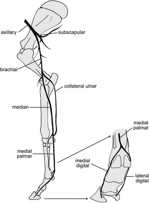
The right and left subclavian arteries follow the same course on each side of the body and each gives off similar branches. Within the thorax each subclavian artery gives off a number of branches that supply blood to the caudal part of the neck, much of the thoracic wall, and the dorsal part of the shoulder. The subclavian artery passes cranial to the first rib on the respective side, passing into the axilla (armpit) of the thoracic limb, where it is called the axillary artery. The axillary artery enters the limb, becoming the brachial artery in the region of the brachium and then the median artery as it continues distal to the elbow. The largest terminal branch of the median artery in the horse is the medial palmar artery, which passes distad in the metacarpus to the fetlock, where it divides into medial and lateral digital arteries. in ruminants, the median artery is continued in the manus as the palmar common digital artery (Fig. 17-8).
Arterial Distribution to the Pelvic Limb
The abdominal aorta terminates near the lumbosacral junction in the two internal iliac arteries (and often a small, midline continuation called the median sacral artery). Each internal iliac artery and its many branches supply the region of the pelvis, the hip, and much of the genitalia.
Just cranial to the internal iliac arteries, the external iliac arteries arise and give rise to branches serving caudoventral parts of the abdominal wall and structures of the inguinal region (prepuce, scrotum, and/or mammary gland).
These large arteries then continue into the pelvic limbs as the femoral arteries. The femoral artery descends on the medial aspect of the limb, giving branches to the large thigh muscles, and continues in the region of the caudal stifle as the popliteal artery. After a very short course, the popliteal artery divides into cranial and caudal tibial arteries. The small caudal tibial artery supplies the muscles of the crus, or true leg. The cranial tibial artery is larger; it passes craniad between the tibia and fibula and descends on the cranial side of the crus to the hock. Where this vessel lies on the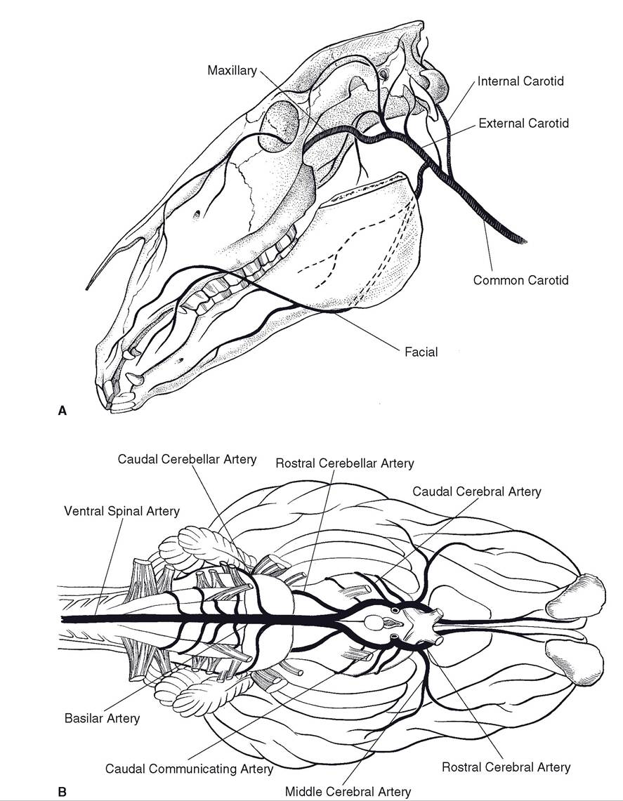
Figure 17-7. Blood supply to the head and brain. A) The common carotid artery branches into a large external carotid artery supplying most of the head and the internal carotid artery, which enters the skull to supply the brain. B) Ventral view of the brain. Internal carotid arteries enter the cerebral arterial circle.
Figure 17-8. Blood supply to the equine thoracic limb, medial view.
flexor surface of the hock, it is referred to as the dorsal pedal artery. In horses, it continues distad as the dorsal (great) metatarsal a. III, running on the lateral side of the pes in the groove between the cannon bone and lateral splint. ultimately, it passes to the plantar aspect of the distal cannon bone by crossing deep to the splint bone. At the equine fetlock it divides into medial and lateral digital arteries. In ruminants, the dorsal pedal artery continues distad on the dorsal aspect of the pes; the plantar side is supplied by a continuation of the saphenous artery, a medial branch of the femoral artery (Fig. 17-9).